 Movie
Movie Controller

[English] 日本語
 Yorodumi
- PDB-4fi5: Crystal structure of the N-terminal domain of Hantaan virus strai... -
+ Open data

- Basic information
| Entry | Database: PDB / ID: 4fi5 | ||||||
|---|---|---|---|---|---|---|---|
| Title | Crystal structure of the N-terminal domain of Hantaan virus strain 76-118 nucleoprotein | ||||||
 Components Components | Nucleoprotein | ||||||
 Keywords Keywords | VIRAL PROTEIN / Structural Genomics / NIAID / National Institute of Allergy and Infectious Diseases / Seattle Structural Genomics Center for Infectious Disease / SSGCID / Korean hemorrhagic fever virus / Hantaan virus / Hantavirus / NP / nucleoprotein / antibody epitope / N-terminal domain / ssRNA negative strand virus / Bunyaviridae / human host / Eurasian field mouse host / virion | ||||||
| Function / homology |  Function and homology information Function and homology information: / helical viral capsid / : / host cell Golgi apparatus / viral nucleocapsid / endonuclease activity / Hydrolases; Acting on ester bonds / host cell perinuclear region of cytoplasm / ribonucleoprotein complex / RNA bindingSimilarity search - Function | ||||||
| Biological species |  Hantaan virus Hantaan virus | ||||||
| Method | X-RAY DIFFRACTION / SYNCHROTRON / MOLECULAR REPLACEMENT / molecular replacement / Resolution: 2.2 Å | ||||||
 Authors Authors | Seattle Structural Genomics Center for Infectious Disease (SSGCID) | ||||||
 Citation Citation | Journal: TO BE PUBLISHED Title: Crystal structure of the N-terminal domain of Hantaan virus strain 76-118 nucleoprotein Authors: Edwards, T.E. / Abendroth, J. / Altamura, L. / Seattle Structural Genomics Center for Infectious Disease (SSGCID) | ||||||
| History |
|
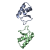
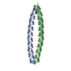
- Structure visualization
| Structure viewer | Molecule: MolmilJmol/JSmol |
|---|
- Downloads & links
-Download
| PDBx/mmCIF format | 4fi5.cif.gz | 43.9 KB | Display | PDBx/mmCIF format |
|---|---|---|---|---|
| PDB format | pdb4fi5.ent.gz | 29.8 KB | Display | PDB format |
| PDBx/mmJSON format | 4fi5.json.gz | Tree view | PDBx/mmJSON format | |
| Others |  Other downloads Other downloads |
-Validation report
| Arichive directory | https://data.pdbj.org/pub/pdb/validation_reports/fi/4fi5ftp://data.pdbj.org/pub/pdb/validation_reports/fi/4fi5 | HTTPS FTP |
|---|
-Related structure data
| Related structure data | 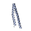 2ic9S S: Starting model for refinement |
|---|---|
| Similar structure data | |
| Other databases |


-Links
 PDBj
PDBj- Assembly
| Deposited unit | 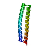
| ||||||||
|---|---|---|---|---|---|---|---|---|---|
| 1 |
| ||||||||
| Unit cell |
|
-Components
| #1: Protein | / Nucleocapsid protein / Protein N Mass: 12829.333 Da / Num. of mol.: 1 / Fragment: unp residues 3-93 Source method: isolated from a genetically manipulated source Source: (gene. exp.) Hantaan virus / Strain: 76-118 / Gene: N / Plasmid: pAVA0421 / Production host:  Escherichia coli (E. coli) / References: UniProt: P05133 Escherichia coli (E. coli) / References: UniProt: P05133 |
|---|---|
| #2: Water | ChemComp-HOH / Water Mass: 18.015 Da / Num. of mol.: 71 / Source method: isolated from a natural source / Formula: H2O Mass: 18.015 Da / Num. of mol.: 71 / Source method: isolated from a natural source / Formula: H2O |
-Experimental details
-Experiment
| Experiment | Method: X-RAY DIFFRACTION / Number of used crystals: 1 |
|---|
- Sample preparation
| Crystal | Density Matthews: 2.46 Å3/Da / Density % sol: 50.03 % |
|---|---|
| Crystal grow | Temperature: 289 K / Method: vapor diffusion, sitting drop / pH: 7.5 Details: HahaA.17785.a.A16.PS01490 at 20 mg/mL against Morpheus screen condition g8, 12.5% PEG1000, 12.5% PEG3350, 12.5% MPD, 20 mM Na-formate, 20 mM Na-citrate, 20 mM Ammonium acetate, 20 mM NaK ...Details: HahaA.17785.a.A16.PS01490 at 20 mg/mL against Morpheus screen condition g8, 12.5% PEG1000, 12.5% PEG3350, 12.5% MPD, 20 mM Na-formate, 20 mM Na-citrate, 20 mM Ammonium acetate, 20 mM NaK tartrate, 100 mM MOPS/HEPES-Na pH 7.5, crystal tracking ID 234641g8, puck ID hky1-5, VAPOR DIFFUSION, SITTING DROP, temperature 289K |
-Data collection
| Diffraction | Mean temperature: 100 K | |||||||||||||||||||||||||||||||||||||||||||||||||||||||||||||||||||||||||||||||||||||||||||||||||||||||||||||||||||||||||||||||||||||||||||||||||||
|---|---|---|---|---|---|---|---|---|---|---|---|---|---|---|---|---|---|---|---|---|---|---|---|---|---|---|---|---|---|---|---|---|---|---|---|---|---|---|---|---|---|---|---|---|---|---|---|---|---|---|---|---|---|---|---|---|---|---|---|---|---|---|---|---|---|---|---|---|---|---|---|---|---|---|---|---|---|---|---|---|---|---|---|---|---|---|---|---|---|---|---|---|---|---|---|---|---|---|---|---|---|---|---|---|---|---|---|---|---|---|---|---|---|---|---|---|---|---|---|---|---|---|---|---|---|---|---|---|---|---|---|---|---|---|---|---|---|---|---|---|---|---|---|---|---|---|---|---|
| Diffraction source | Source: SYNCHROTRON / Site: SSRL  / Beamline: BL7-1 / Wavelength: 1.003317 Å / Beamline: BL7-1 / Wavelength: 1.003317 Å | |||||||||||||||||||||||||||||||||||||||||||||||||||||||||||||||||||||||||||||||||||||||||||||||||||||||||||||||||||||||||||||||||||||||||||||||||||
| Detector | Type: ADSC QUANTUM 315r / Detector: CCD / Date: May 31, 2012 | |||||||||||||||||||||||||||||||||||||||||||||||||||||||||||||||||||||||||||||||||||||||||||||||||||||||||||||||||||||||||||||||||||||||||||||||||||
| Radiation | Protocol: SINGLE WAVELENGTH / Monochromatic (M) / Laue (L): M / Scattering type: x-ray | |||||||||||||||||||||||||||||||||||||||||||||||||||||||||||||||||||||||||||||||||||||||||||||||||||||||||||||||||||||||||||||||||||||||||||||||||||
| Radiation wavelength | Wavelength: 1.003317 Å / Relative weight: 1 | |||||||||||||||||||||||||||||||||||||||||||||||||||||||||||||||||||||||||||||||||||||||||||||||||||||||||||||||||||||||||||||||||||||||||||||||||||
| Reflection | Resolution: 2.2→50 Å / Num. all: 6619 / Num. obs: 6604 / % possible obs: 99.8 % / Observed criterion σ(I): -3 / Redundancy: 7.3 % / Biso Wilson estimate: 37.089 Å2 / Rmerge(I) obs: 0.078 / Net I/σ(I): 21.38 | |||||||||||||||||||||||||||||||||||||||||||||||||||||||||||||||||||||||||||||||||||||||||||||||||||||||||||||||||||||||||||||||||||||||||||||||||||
| Reflection shell | Diffraction-ID: 1
|
-Phasing
| Phasing | Method: molecular replacement | |||||||||
|---|---|---|---|---|---|---|---|---|---|---|
| Phasing MR | Model details: Phaser MODE: MR_AUTO
|
- Processing
| Software |
| ||||||||||||||||||||||||||||||||||||||||||||||||||||||||||||
|---|---|---|---|---|---|---|---|---|---|---|---|---|---|---|---|---|---|---|---|---|---|---|---|---|---|---|---|---|---|---|---|---|---|---|---|---|---|---|---|---|---|---|---|---|---|---|---|---|---|---|---|---|---|---|---|---|---|---|---|---|---|
| Refinement | Method to determine structure: MOLECULAR REPLACEMENT Starting model: pdb ID 2ic9 Resolution: 2.2→50 Å / Cor.coef. Fo:Fc: 0.952 / Cor.coef. Fo:Fc free: 0.932 / WRfactor Rfree: 0.2087 / WRfactor Rwork: 0.1708 / Occupancy max: 1 / Occupancy min: 0.5 / FOM work R set: 0.8625 / SU B: 7.962 / SU ML: 0.11 / SU R Cruickshank DPI: 0.191 / SU Rfree: 0.1766 / Cross valid method: THROUGHOUT / σ(F): 0 / ESU R: 0.191 / ESU R Free: 0.177 / Stereochemistry target values: MAXIMUM LIKELIHOOD Details: U VALUES : WITH TLS ADDED HYDROGENS HAVE BEEN ADDED IN THE RIDING POSITIONS
| ||||||||||||||||||||||||||||||||||||||||||||||||||||||||||||
| Solvent computation | Ion probe radii: 0.8 Å / Shrinkage radii: 0.8 Å / VDW probe radii: 1.2 Å / Solvent model: MASK | ||||||||||||||||||||||||||||||||||||||||||||||||||||||||||||
| Displacement parameters | Biso max: 73.23 Å2 / Biso mean: 35.4455 Å2 / Biso min: 18.61 Å2
| ||||||||||||||||||||||||||||||||||||||||||||||||||||||||||||
| Refinement step | Cycle: LAST / Resolution: 2.2→50 Å
| ||||||||||||||||||||||||||||||||||||||||||||||||||||||||||||
| Refine LS restraints |
| ||||||||||||||||||||||||||||||||||||||||||||||||||||||||||||
| LS refinement shell | Resolution: 2.2→2.257 Å / Total num. of bins used: 20
| ||||||||||||||||||||||||||||||||||||||||||||||||||||||||||||
| Refinement TLS params. | Method: refined / Origin x: 30.1016 Å / Origin y: 12.5975 Å / Origin z: -0.2086 Å
| ||||||||||||||||||||||||||||||||||||||||||||||||||||||||||||
| Refinement TLS group |
|
