 Movie
Movie Controller

[English] 日本語
 Yorodumi
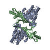
- PDB-4fgm: Crystal structure of the aminopeptidase N family protein Q5QTY1 f... -
+ Open data

- Basic information
| Entry | Database: PDB / ID: 4fgm | ||||||
|---|---|---|---|---|---|---|---|
| Title | Crystal structure of the aminopeptidase N family protein Q5QTY1 from Idiomarina loihiensis. Northeast Structural Genomics Consortium Target IlR60. | ||||||
 Components Components | Aminopeptidase N family protein | ||||||
 Keywords Keywords |  HYDROLASE / Structural Genomics / PSI-Biology / Northeast Structural Genomics Consortium / NESG / peptidase_M61 / PDZ / PDZ_2 HYDROLASE / Structural Genomics / PSI-Biology / Northeast Structural Genomics Consortium / NESG / peptidase_M61 / PDZ / PDZ_2 | ||||||
| Function / homology |  Function and homology information Function and homology information | ||||||
| Biological species |  Idiomarina loihiensis L2TR (bacteria) Idiomarina loihiensis L2TR (bacteria) | ||||||
| Method | X-RAY DIFFRACTION / SYNCHROTRON / SAD / Resolution: 2.394 Å | ||||||
 Authors Authors | Vorobiev, S. / Su, M. / Tong, T. / Kohan, E. / Wang, D. / Everett, J.K. / Acton, T.B. / Montelione, G.T. / Tong, L. / Hunt, J.F. / Northeast Structural Genomics Consortium (NESG) | ||||||
 Citation Citation | Journal: To be Published Title: Crystal structure of the aminopeptidase N family protein Q5QTY1 from Idiomarina loihiensis. Authors: Vorobiev, S. / Su, M. / Tong, T. / Kohan, E. / Wang, D. / Everett, J.K. / Acton, T.B. / Montelione, G.T. / Tong, L. / Hunt, J.F. | ||||||
| History |
|
- Structure visualization
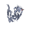
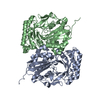
| Structure viewer | Molecule: MolmilJmol/JSmol |
|---|
- Downloads & links
-Download
| PDBx/mmCIF format | 4fgm.cif.gz | 243.9 KB | Display | PDBx/mmCIF format |
|---|---|---|---|---|
| PDB format | pdb4fgm.ent.gz | 204.6 KB | Display | PDB format |
| PDBx/mmJSON format | 4fgm.json.gz | Tree view | PDBx/mmJSON format | |
| Others |  Other downloads Other downloads |
-Validation report
| Arichive directory | https://data.pdbj.org/pub/pdb/validation_reports/fg/4fgmftp://data.pdbj.org/pub/pdb/validation_reports/fg/4fgm | HTTPS FTP |
|---|
-Related structure data
| Similar structure data | |
|---|---|
| Other databases |


-Links
 PDBj
PDBj
- Assembly
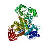
| Deposited unit | 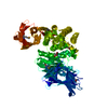
| ||||||||
|---|---|---|---|---|---|---|---|---|---|
| 1 |
| ||||||||
| 2 | 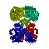
| ||||||||
| Unit cell |
| ||||||||
| Details | monomer according to aggregation screening |
-Components
| #1: Protein | Mass: 68525.758 Da / Num. of mol.: 1 Source method: isolated from a genetically manipulated source Source: (gene. exp.) Idiomarina loihiensis L2TR (bacteria) / Strain: ATCC BAA-735 / DSM 15497 / L2-TR / Gene: IL1258 / Plasmid: pET21_NESG, ILR60-21.5 / Production host: Escherichia coli (E. coli) / Strain (production host): BL21(DE3)+Magic / References: UniProt: Q5QTY1, membrane alanyl aminopeptidase |
|---|---|
| #2: Chemical | ChemComp-MAE / Maleic acid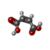  Mass: 116.072 Da / Num. of mol.: 1 / Source method: obtained synthetically / Formula: C4H4O4 Mass: 116.072 Da / Num. of mol.: 1 / Source method: obtained synthetically / Formula: C4H4O4 |
| #3: Chemical | ChemComp-ZN /   Mass: 65.409 Da / Num. of mol.: 1 / Source method: obtained synthetically / Formula: Zn Mass: 65.409 Da / Num. of mol.: 1 / Source method: obtained synthetically / Formula: Zn |
| #4: Water | ChemComp-HOH / Water Mass: 18.015 Da / Num. of mol.: 222 / Source method: isolated from a natural source / Formula: H2O Mass: 18.015 Da / Num. of mol.: 222 / Source method: isolated from a natural source / Formula: H2O |
-Experimental details
-Experiment
| Experiment | Method: X-RAY DIFFRACTION / Number of used crystals: 1 |
|---|
- Sample preparation
| Crystal | Density Matthews: 3.32 Å3/Da / Density % sol: 62.94 % |
|---|---|
| Crystal grow | Temperature: 277 K / Method: microbatch crystallization under oil / pH: 6.8 Details: Protein solution: 100mM NaCl, 5mM DTT, 0.02% NaN3, 10mM Tris-HCl (pH 7.5). Reservoir solution: 45% PEG/Tacsimate pH 6.8 (Hampton research HR2-092), 45% Silver Bullet (Hampton research HR2- ...Details: Protein solution: 100mM NaCl, 5mM DTT, 0.02% NaN3, 10mM Tris-HCl (pH 7.5). Reservoir solution: 45% PEG/Tacsimate pH 6.8 (Hampton research HR2-092), 45% Silver Bullet (Hampton research HR2-096, G11), 0.1 M sodium citrate., Microbatch crystallization under oil, temperature 277K |
-Data collection
| Diffraction | Mean temperature: 100 K |
|---|---|
| Diffraction source | Source: SYNCHROTRON / Site: SSRL  / Beamline: BL9-2 / Wavelength: 0.979 Å / Beamline: BL9-2 / Wavelength: 0.979 Å |
| Detector | Type: MARMOSAIC 325 mm CCD / Detector: CCD / Date: May 20, 2012 |
| Radiation | Protocol: SINGLE WAVELENGTH / Monochromatic (M) / Laue (L): M / Scattering type: x-ray |
| Radiation wavelength | Wavelength: 0.979 Å / Relative weight: 1 |
| Reflection | Resolution: 2.394→50 Å / % possible obs: 100 % / Observed criterion σ(F): 0 / Observed criterion σ(I): 0 / Redundancy: 11.7 % / Biso Wilson estimate: 49.99 Å2 / Rmerge(I) obs: 0.115 / Net I/σ(I): 27.3 |
| Reflection shell | Resolution: 2.4→2.49 Å / Redundancy: 11.7 % / Rmerge(I) obs: 0.978 / Mean I/σ(I) obs: 2.05 / Num. unique all: 27765 / % possible all: 100 |
- Processing
| Software |
| ||||||||||||||||||||||||||||||||||||||||||||||||||||||||||||||||||||||||||||||||||||||||||||||||||||||||||||||||||||||||||||||||||||||||||||||||||||||||||||
|---|---|---|---|---|---|---|---|---|---|---|---|---|---|---|---|---|---|---|---|---|---|---|---|---|---|---|---|---|---|---|---|---|---|---|---|---|---|---|---|---|---|---|---|---|---|---|---|---|---|---|---|---|---|---|---|---|---|---|---|---|---|---|---|---|---|---|---|---|---|---|---|---|---|---|---|---|---|---|---|---|---|---|---|---|---|---|---|---|---|---|---|---|---|---|---|---|---|---|---|---|---|---|---|---|---|---|---|---|---|---|---|---|---|---|---|---|---|---|---|---|---|---|---|---|---|---|---|---|---|---|---|---|---|---|---|---|---|---|---|---|---|---|---|---|---|---|---|---|---|---|---|---|---|---|---|---|---|
| Refinement | Method to determine structure: SAD / Resolution: 2.394→42.325 Å / Occupancy max: 1 / Occupancy min: 1 / SU ML: 0.68 / Cross valid method: THROUGHOUT / σ(F): 1.38 / Phase error: 20.6 / Stereochemistry target values: ML
| ||||||||||||||||||||||||||||||||||||||||||||||||||||||||||||||||||||||||||||||||||||||||||||||||||||||||||||||||||||||||||||||||||||||||||||||||||||||||||||
| Solvent computation | Shrinkage radii: 0.98 Å / VDW probe radii: 1.2 Å / Solvent model: FLAT BULK SOLVENT MODEL / Bsol: 40.973 Å2 / ksol: 0.326 e/Å3 | ||||||||||||||||||||||||||||||||||||||||||||||||||||||||||||||||||||||||||||||||||||||||||||||||||||||||||||||||||||||||||||||||||||||||||||||||||||||||||||
| Displacement parameters | Biso max: 144.29 Å2 / Biso mean: 51.705 Å2 / Biso min: 24.74 Å2
| ||||||||||||||||||||||||||||||||||||||||||||||||||||||||||||||||||||||||||||||||||||||||||||||||||||||||||||||||||||||||||||||||||||||||||||||||||||||||||||
| Refinement step | Cycle: LAST / Resolution: 2.394→42.325 Å
| ||||||||||||||||||||||||||||||||||||||||||||||||||||||||||||||||||||||||||||||||||||||||||||||||||||||||||||||||||||||||||||||||||||||||||||||||||||||||||||
| Refine LS restraints |
| ||||||||||||||||||||||||||||||||||||||||||||||||||||||||||||||||||||||||||||||||||||||||||||||||||||||||||||||||||||||||||||||||||||||||||||||||||||||||||||
| LS refinement shell | Refine-ID: X-RAY DIFFRACTION / Total num. of bins used: 25 / % reflection obs: 100 %
|
