 Movie
Movie Controller

[English] 日本語
 Yorodumi
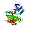
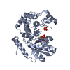
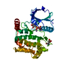
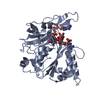
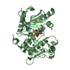
- PDB-4f1o: Crystal Structure of the L1180T mutant Roco4 Kinase Domain from D... -
+ Open data

- Basic information
| Entry | Database: PDB / ID: 4f1o | ||||||
|---|---|---|---|---|---|---|---|
| Title | Crystal Structure of the L1180T mutant Roco4 Kinase Domain from D. discoideum bound to AppCp | ||||||
 Components Components | Serine/threonine-protein kinase roco4 | ||||||
 Keywords Keywords |  Transferase / Signaling Protein / Protein Kinase / LRRK2 / ROCO / Kinase / ATP-binding / Nucleotide-binding / Serine/threonine-protein kinase Transferase / Signaling Protein / Protein Kinase / LRRK2 / ROCO / Kinase / ATP-binding / Nucleotide-binding / Serine/threonine-protein kinase | ||||||
| Function / homology |  Function and homology information Function and homology informationsorocarp stalk development / Regulation of necroptotic cell death / phototaxis / mitochondrial electron transport, NADH to ubiquinone / response to reactive oxygen species / protein tyrosine kinase activity / non-specific serine/threonine protein kinase / protein kinase activity / phosphorylation / protein serine kinase activity ...sorocarp stalk development / Regulation of necroptotic cell death / phototaxis / mitochondrial electron transport, NADH to ubiquinone / response to reactive oxygen species / protein tyrosine kinase activity / non-specific serine/threonine protein kinase / protein kinase activity / phosphorylation / protein serine kinase activity / protein serine/threonine kinase activity / GTP binding / signal transduction / mitochondrion / ATP binding / cytosol / cytoplasmSimilarity search - Function | ||||||
| Biological species |  Dictyostelium discoideum (eukaryote) Dictyostelium discoideum (eukaryote) | ||||||
| Method | X-RAY DIFFRACTION / SYNCHROTRON / MOLECULAR REPLACEMENT / Resolution: 2.3 Å | ||||||
 Authors Authors | Gilsbach, B.K. / Vetter, I.R. / Wittinghofer, A. / Kortholt, A. | ||||||
 Citation Citation | Journal: Proc.Natl.Acad.Sci.USA / Year: 2012 Title: Roco kinase structures give insights into the mechanism of Parkinson disease-related leucine-rich-repeat kinase 2 mutations. Authors: Gilsbach, B.K. / Ho, F.Y. / Vetter, I.R. / van Haastert, P.J. / Wittinghofer, A. / Kortholt, A. | ||||||
| History |
|
- Structure visualization
| Structure viewer | Molecule: MolmilJmol/JSmol |
|---|
- Downloads & links
-Download
| PDBx/mmCIF format | 4f1o.cif.gz | 75.1 KB | Display | PDBx/mmCIF format |
|---|---|---|---|---|
| PDB format | pdb4f1o.ent.gz | 53.9 KB | Display | PDB format |
| PDBx/mmJSON format | 4f1o.json.gz | Tree view | PDBx/mmJSON format | |
| Others |  Other downloads Other downloads |
-Validation report
| Arichive directory | https://data.pdbj.org/pub/pdb/validation_reports/f1/4f1oftp://data.pdbj.org/pub/pdb/validation_reports/f1/4f1o | HTTPS FTP |
|---|
-Related structure data
| Related structure data |  4f0fC  4f0gC  4f1mC  4f1tC 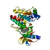 3dtcS C: citing same article ( S: Starting model for refinement |
|---|---|
| Similar structure data |


-Links
 PDBj
PDBj


- Assembly
| Deposited unit | 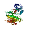
| ||||||||
|---|---|---|---|---|---|---|---|---|---|
| 1 |
| ||||||||
| Unit cell |
| ||||||||
| Components on special symmetry positions |
|
-Components
| #1: Protein | Mass: 32604.484 Da / Num. of mol.: 1 / Fragment: Roco4 Kinase Domain, UNP residues 1019-1292 Source method: isolated from a genetically manipulated source Source: (gene. exp.) Dictyostelium discoideum (eukaryote) / Strain: AX3 / Gene: DDB_G0288251, roco4 / Plasmid: pGEX4T1 NTev / Production host:  Escherichia coli (E. coli) / Strain (production host): BL21(DE3) / References: UniProt: Q6XHB2 Escherichia coli (E. coli) / Strain (production host): BL21(DE3) / References: UniProt: Q6XHB2 |
|---|---|
| #2: Chemical | ChemComp-ACP /   Mass: 505.208 Da / Num. of mol.: 1 / Source method: obtained synthetically / Formula: C11H18N5O12P3 / Comment: AMP-PCP, energy-carrying molecule analogue*YM Mass: 505.208 Da / Num. of mol.: 1 / Source method: obtained synthetically / Formula: C11H18N5O12P3 / Comment: AMP-PCP, energy-carrying molecule analogue*YM |
| #3: Water | ChemComp-HOH / Water Mass: 18.015 Da / Num. of mol.: 165 / Source method: isolated from a natural source / Formula: H2O Mass: 18.015 Da / Num. of mol.: 165 / Source method: isolated from a natural source / Formula: H2O |
-Experimental details
-Experiment
| Experiment | Method: X-RAY DIFFRACTION / Number of used crystals: 1 |
|---|
- Sample preparation
| Crystal | Density Matthews: 2.29 Å3/Da / Density % sol: 46.37 % |
|---|---|
| Crystal grow | Temperature: 277 K / Method: vapor diffusion, hanging drop / pH: 8.5 Details: 0.1 M Bis- Tris, 0.2 M Na/K Tartrate; 11% PEG 3350, pH 8.5, vapor diffusion, hanging drop, temperature 277K |
-Data collection
| Diffraction | Mean temperature: 100 K | ||||||||||||||||||||||||||||||||||||||||||||||||||||||||||||||||||||||||||||||||||||||||||||||||||||||||||||||||||||||||||||||
|---|---|---|---|---|---|---|---|---|---|---|---|---|---|---|---|---|---|---|---|---|---|---|---|---|---|---|---|---|---|---|---|---|---|---|---|---|---|---|---|---|---|---|---|---|---|---|---|---|---|---|---|---|---|---|---|---|---|---|---|---|---|---|---|---|---|---|---|---|---|---|---|---|---|---|---|---|---|---|---|---|---|---|---|---|---|---|---|---|---|---|---|---|---|---|---|---|---|---|---|---|---|---|---|---|---|---|---|---|---|---|---|---|---|---|---|---|---|---|---|---|---|---|---|---|---|---|---|
| Diffraction source | Source: SYNCHROTRON / Site: SLS  / Beamline: X10SA / Wavelength: 0.9778 Å / Beamline: X10SA / Wavelength: 0.9778 Å | ||||||||||||||||||||||||||||||||||||||||||||||||||||||||||||||||||||||||||||||||||||||||||||||||||||||||||||||||||||||||||||||
| Detector | Type: DECTRIS PILATUS 6M / Detector: PIXEL / Date: Mar 20, 2011 | ||||||||||||||||||||||||||||||||||||||||||||||||||||||||||||||||||||||||||||||||||||||||||||||||||||||||||||||||||||||||||||||
| Radiation | Monochromator: SAGITALLY - HORIZONTALLY FOCUSED SI(111) MONOCHROMATOR Protocol: SINGLE WAVELENGTH / Monochromatic (M) / Laue (L): M / Scattering type: x-ray | ||||||||||||||||||||||||||||||||||||||||||||||||||||||||||||||||||||||||||||||||||||||||||||||||||||||||||||||||||||||||||||||
| Radiation wavelength | Wavelength: 0.9778 Å / Relative weight: 1 | ||||||||||||||||||||||||||||||||||||||||||||||||||||||||||||||||||||||||||||||||||||||||||||||||||||||||||||||||||||||||||||||
| Reflection | Resolution: 2.3→42 Å / Num. all: 14611 / Num. obs: 14604 / % possible obs: 100 % / Observed criterion σ(F): -3 / Observed criterion σ(I): -3 / Rmerge(I) obs: 0.219 / Net I/σ(I): 12.09 | ||||||||||||||||||||||||||||||||||||||||||||||||||||||||||||||||||||||||||||||||||||||||||||||||||||||||||||||||||||||||||||||
| Reflection shell |
|
- Processing
| Software |
| ||||||||||||||||||||||||||||||||||||||||||||||||||||||||||||||||||||||||||||||||||||||||||||||||||||||||||||||||||||||||||||||||||||||||||||||||||||||||||||||||||||||||||
|---|---|---|---|---|---|---|---|---|---|---|---|---|---|---|---|---|---|---|---|---|---|---|---|---|---|---|---|---|---|---|---|---|---|---|---|---|---|---|---|---|---|---|---|---|---|---|---|---|---|---|---|---|---|---|---|---|---|---|---|---|---|---|---|---|---|---|---|---|---|---|---|---|---|---|---|---|---|---|---|---|---|---|---|---|---|---|---|---|---|---|---|---|---|---|---|---|---|---|---|---|---|---|---|---|---|---|---|---|---|---|---|---|---|---|---|---|---|---|---|---|---|---|---|---|---|---|---|---|---|---|---|---|---|---|---|---|---|---|---|---|---|---|---|---|---|---|---|---|---|---|---|---|---|---|---|---|---|---|---|---|---|---|---|---|---|---|---|---|---|---|---|
| Refinement | Method to determine structure: MOLECULAR REPLACEMENT Starting model: 3DTC Resolution: 2.3→41.98 Å / Cor.coef. Fo:Fc: 0.895 / Cor.coef. Fo:Fc free: 0.856 / Occupancy max: 1 / Occupancy min: 0.5 / SU B: 8.735 / SU ML: 0.214 / Cross valid method: THROUGHOUT / ESU R: 0.441 / ESU R Free: 0.289 / Stereochemistry target values: MAXIMUM LIKELIHOOD / Details: HYDROGENS HAVE BEEN USED IF PRESENT IN THE INPUT
| ||||||||||||||||||||||||||||||||||||||||||||||||||||||||||||||||||||||||||||||||||||||||||||||||||||||||||||||||||||||||||||||||||||||||||||||||||||||||||||||||||||||||||
| Solvent computation | Ion probe radii: 0.8 Å / Shrinkage radii: 0.8 Å / VDW probe radii: 1.2 Å / Solvent model: MASK | ||||||||||||||||||||||||||||||||||||||||||||||||||||||||||||||||||||||||||||||||||||||||||||||||||||||||||||||||||||||||||||||||||||||||||||||||||||||||||||||||||||||||||
| Displacement parameters | Biso mean: 33.167 Å2
| ||||||||||||||||||||||||||||||||||||||||||||||||||||||||||||||||||||||||||||||||||||||||||||||||||||||||||||||||||||||||||||||||||||||||||||||||||||||||||||||||||||||||||
| Refinement step | Cycle: LAST / Resolution: 2.3→41.98 Å
| ||||||||||||||||||||||||||||||||||||||||||||||||||||||||||||||||||||||||||||||||||||||||||||||||||||||||||||||||||||||||||||||||||||||||||||||||||||||||||||||||||||||||||
| Refine LS restraints |
| ||||||||||||||||||||||||||||||||||||||||||||||||||||||||||||||||||||||||||||||||||||||||||||||||||||||||||||||||||||||||||||||||||||||||||||||||||||||||||||||||||||||||||
| LS refinement shell | Resolution: 2.3→2.36 Å / Total num. of bins used: 20
|
