 Movie
Movie Controller

[English] 日本語
 Yorodumi
- PDB-4f1j: Crystal structure of the MG2+ loaded VWA domain of plasmodium fal... -
+ Open data

- Basic information
| Entry | Database: PDB / ID: 4f1j | ||||||
|---|---|---|---|---|---|---|---|
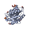
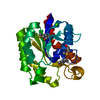
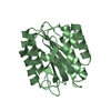
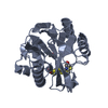
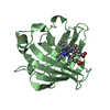
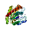
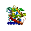
| Title | Crystal structure of the MG2+ loaded VWA domain of plasmodium falciparum trap protein | ||||||
 Components Components | Thrombospondin related anonymous protein | ||||||
 Keywords Keywords |  CELL ADHESION / Rossmann fold / DINUCLEOTIDE BINDING / MEMBRANE CELL ADHESION / Rossmann fold / DINUCLEOTIDE BINDING / MEMBRANE | ||||||
| Function / homology |  Function and homology information Function and homology information | ||||||
| Biological species |  Plasmodium falciparum (malaria parasite P. falciparum) Plasmodium falciparum (malaria parasite P. falciparum) | ||||||
| Method | X-RAY DIFFRACTION / SYNCHROTRON / SIRAS / Resolution: 1.73 Å | ||||||
 Authors Authors | Pihlajamaa, T. / Knuuti, J. / Kajander, T. / Sharma, A. / Permi, P. | ||||||
 Citation Citation | Journal: Biochem.J. / Year: 2013 Title: Structure of Plasmodium falciparum TRAP (thrombospondin-related anonymous protein) A domain highlights distinct features in apicomplexan von Willebrand factor A homologues. Authors: Pihlajamaa, T. / Kajander, T. / Knuuti, J. / Horkka, K. / Sharma, A. / Permi, P. | ||||||
| History |
|
- Structure visualization
| Structure viewer | Molecule: MolmilJmol/JSmol |
|---|
- Downloads & links
-Download
| PDBx/mmCIF format | 4f1j.cif.gz | 99.8 KB | Display | PDBx/mmCIF format |
|---|---|---|---|---|
| PDB format | pdb4f1j.ent.gz | 80.7 KB | Display | PDB format |
| PDBx/mmJSON format | 4f1j.json.gz | Tree view | PDBx/mmJSON format | |
| Others |  Other downloads Other downloads |
-Validation report
| Arichive directory | https://data.pdbj.org/pub/pdb/validation_reports/f1/4f1jftp://data.pdbj.org/pub/pdb/validation_reports/f1/4f1j | HTTPS FTP |
|---|
-Related structure data


-Links
 PDBj
PDBj


- Assembly
| Deposited unit | 
| |||||||||||||||||||||||||||
|---|---|---|---|---|---|---|---|---|---|---|---|---|---|---|---|---|---|---|---|---|---|---|---|---|---|---|---|---|
| 1 | 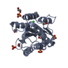
| |||||||||||||||||||||||||||
| 2 | 
| |||||||||||||||||||||||||||
| Unit cell |
| |||||||||||||||||||||||||||
| Noncrystallographic symmetry (NCS) | NCS domain:
NCS domain segments:
|
-Components
-Protein , 1 types, 2 molecules AB
| #1: Protein | Mass: 22766.244 Da / Num. of mol.: 2 Source method: isolated from a genetically manipulated source Source: (gene. exp.) Plasmodium falciparum (malaria parasite P. falciparum)Gene: THROMBOSPONDIN RELATED ANONYMOUS PROTEIN (TRAP), TRAP / Plasmid: pRAT4 / Production host:  Escherichia coli (E. coli) / Strain (production host): BL21(DE3) / References: UniProt: Q01502 Escherichia coli (E. coli) / Strain (production host): BL21(DE3) / References: UniProt: Q01502 |
|---|
-Non-polymers , 5 types, 416 molecules 


| #2: Chemical | ChemComp-MG /  Mass: 24.305 Da / Num. of mol.: 4 / Source method: obtained synthetically / Formula: Mg Mass: 24.305 Da / Num. of mol.: 4 / Source method: obtained synthetically / Formula: Mg#3: Chemical | ChemComp-SO4 / Sulfate Mass: 96.063 Da / Num. of mol.: 7 / Source method: obtained synthetically / Formula: SO4 Mass: 96.063 Da / Num. of mol.: 7 / Source method: obtained synthetically / Formula: SO4#4: Chemical | ChemComp-EDO / Ethylene glycol Mass: 62.068 Da / Num. of mol.: 12 / Source method: obtained synthetically / Formula: C2H6O2 Mass: 62.068 Da / Num. of mol.: 12 / Source method: obtained synthetically / Formula: C2H6O2#5: Chemical | Chloride Mass: 35.453 Da / Num. of mol.: 2 / Source method: obtained synthetically / Formula: Cl Mass: 35.453 Da / Num. of mol.: 2 / Source method: obtained synthetically / Formula: Cl#6: Water | ChemComp-HOH / | WaterMass: 18.015 Da / Num. of mol.: 391 / Source method: isolated from a natural source / Formula: H2O |
|---|
-Experimental details
-Experiment
| Experiment | Method: X-RAY DIFFRACTION / Number of used crystals: 1 |
|---|
- Sample preparation
| Crystal | Density Matthews: 2.28 Å3/Da / Density % sol: 46.09 % |
|---|---|
| Crystal grow | Temperature: 293 K / Method: vapor diffusion, sitting drop / pH: 5.5 Details: 25% PEG 3350, 0.1M BIS-TRIS, 0.2M MAGNESIUM SULFATE, pH 5.5, VAPOR DIFFUSION, SITTING DROP, temperature 293.0K |
-Data collection
| Diffraction | Mean temperature: 100 K |
|---|---|
| Diffraction source | Source: SYNCHROTRON / Site: SLS  / Beamline: X06DA / Wavelength: 1 Å / Beamline: X06DA / Wavelength: 1 Å |
| Detector | Type: MARMOSAIC 225 mm CCD / Detector: CCD / Date: May 24, 2010 Details: THE OPTICAL ELEMENTS CONSIST OF A VERTICALLY COLLIMATING MIRROR (M1, FOCUS AT INFINITY) FOLLOWED BY A BARTELS MONOCHROMATOR WITH DUAL CHANNEL CUT CRYSTALS (DCCM) IN (+--+) GEOMETRY, AND A ...Details: THE OPTICAL ELEMENTS CONSIST OF A VERTICALLY COLLIMATING MIRROR (M1, FOCUS AT INFINITY) FOLLOWED BY A BARTELS MONOCHROMATOR WITH DUAL CHANNEL CUT CRYSTALS (DCCM) IN (+--+) GEOMETRY, AND A TOROIDAL MIRROR (M2) TO VERTICALLY AND HORIZONTALLY FOCUS THE BEAM AT THE SAMPLE POSITION (WITH 2:1 HORIZONTAL DEMAGNIFICATION) |
| Radiation | Monochromator: BARTELS MONOCHROMATOR / Protocol: SINGLE WAVELENGTH / Monochromatic (M) / Laue (L): M / Scattering type: x-ray |
| Radiation wavelength | Wavelength: 1 Å / Relative weight: 1 |
| Reflection | Resolution: 1.73→50 Å / Num. all: 84445 / % possible obs: 99.8 % / Observed criterion σ(F): -3 / Observed criterion σ(I): -3 / Redundancy: 10.271 % / Rmerge(I) obs: 0.13 / Net I/σ(I): 14.72 |
| Reflection shell | Resolution: 1.73→1.83 Å / Redundancy: 10 % / Rmerge(I) obs: 0.853 / Mean I/σ(I) obs: 2.9 / % possible all: 98.6 |
- Processing
| Software |
| |||||||||||||||||||||||||||||||||||||||||||||||||||||||||||||||||||||||||||||||||||||||||||||||||||||||||||||||||||||||||||||||||||||||
|---|---|---|---|---|---|---|---|---|---|---|---|---|---|---|---|---|---|---|---|---|---|---|---|---|---|---|---|---|---|---|---|---|---|---|---|---|---|---|---|---|---|---|---|---|---|---|---|---|---|---|---|---|---|---|---|---|---|---|---|---|---|---|---|---|---|---|---|---|---|---|---|---|---|---|---|---|---|---|---|---|---|---|---|---|---|---|---|---|---|---|---|---|---|---|---|---|---|---|---|---|---|---|---|---|---|---|---|---|---|---|---|---|---|---|---|---|---|---|---|---|---|---|---|---|---|---|---|---|---|---|---|---|---|---|---|---|
| Refinement | Method to determine structure: SIRAS / Resolution: 1.73→30 Å / Cor.coef. Fo:Fc: 0.961 / Cor.coef. Fo:Fc free: 0.943 / SU B: 2.029 / SU ML: 0.068 / Cross valid method: THROUGHOUT / σ(F): 0 / ESU R: 0.112 / ESU R Free: 0.112 / Stereochemistry target values: MAXIMUM LIKELIHOOD / Details: HYDROGENS HAVE BEEN ADDED IN THE RIDING POSITIONS
| |||||||||||||||||||||||||||||||||||||||||||||||||||||||||||||||||||||||||||||||||||||||||||||||||||||||||||||||||||||||||||||||||||||||
| Solvent computation | Ion probe radii: 0.8 Å / Shrinkage radii: 0.8 Å / VDW probe radii: 1.2 Å / Solvent model: MASK | |||||||||||||||||||||||||||||||||||||||||||||||||||||||||||||||||||||||||||||||||||||||||||||||||||||||||||||||||||||||||||||||||||||||
| Displacement parameters | Biso mean: 11.147 Å2
| |||||||||||||||||||||||||||||||||||||||||||||||||||||||||||||||||||||||||||||||||||||||||||||||||||||||||||||||||||||||||||||||||||||||
| Refinement step | Cycle: LAST / Resolution: 1.73→30 Å
| |||||||||||||||||||||||||||||||||||||||||||||||||||||||||||||||||||||||||||||||||||||||||||||||||||||||||||||||||||||||||||||||||||||||
| Refine LS restraints |
| |||||||||||||||||||||||||||||||||||||||||||||||||||||||||||||||||||||||||||||||||||||||||||||||||||||||||||||||||||||||||||||||||||||||
| Refine LS restraints NCS | Dom-ID: 1 / Auth asym-ID: A / Ens-ID: 1 / Refine-ID: X-RAY DIFFRACTION
| |||||||||||||||||||||||||||||||||||||||||||||||||||||||||||||||||||||||||||||||||||||||||||||||||||||||||||||||||||||||||||||||||||||||
| LS refinement shell | Resolution: 1.73→1.773 Å / Total num. of bins used: 20
|
