 Movie
Movie Controller

[English] 日本語
 Yorodumi
- PDB-4ey8: Crystal structure of recombinant human acetylcholinesterase in co... -
+ Open data

- Basic information
| Entry | Database: PDB / ID: 4ey8 | |||||||||
|---|---|---|---|---|---|---|---|---|---|---|
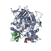
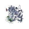
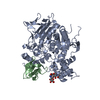
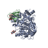
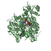
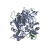
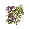
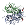
| Title | Crystal structure of recombinant human acetylcholinesterase in complex with fasciculin-2 | |||||||||
 Components Components |
| |||||||||
 Keywords Keywords | HYDROLASE/HYDROLASE INHIBITOR /  acetylcholinesterase / hydrolase / fasciculin 2 / snake venom toxin / inhibitor / HYDROLASE-HYDROLASE INHIBITOR complex acetylcholinesterase / hydrolase / fasciculin 2 / snake venom toxin / inhibitor / HYDROLASE-HYDROLASE INHIBITOR complex | |||||||||
| Function / homology |  Function and homology information Function and homology informationnegative regulation of synaptic transmission, cholinergic / serine hydrolase activity / Neurotransmitter clearance / acetylcholine catabolic process in synaptic cleft / cholinesterase activity / acetylcholine catabolic process / acetylcholine binding / amyloid precursor protein metabolic process / acetylcholinesterase / acetylcholine receptor signaling pathway ...negative regulation of synaptic transmission, cholinergic / serine hydrolase activity / Neurotransmitter clearance / acetylcholine catabolic process in synaptic cleft / cholinesterase activity / acetylcholine catabolic process / acetylcholine binding / amyloid precursor protein metabolic process / acetylcholinesterase / acetylcholine receptor signaling pathway / osteoblast development / acetylcholinesterase activity / choline metabolic process / Synthesis of PC / basement membrane / regulation of receptor recycling / Synthesis, secretion, and deacylation of Ghrelin / laminin binding / side of membrane / synaptic cleft / synapse assembly / collagen binding / positive regulation of protein secretion / neuromuscular junction / receptor internalization / : / retina development in camera-type eye / nervous system development / positive regulation of cold-induced thermogenesis / amyloid-beta binding / toxin activity / hydrolase activity / cell adhesion / synapse / perinuclear region of cytoplasm / Golgi apparatus / cell surface / protein homodimerization activity / extracellular space / extracellular region / membrane / nucleus / plasma membraneSimilarity search - Function | |||||||||
| Biological species |  Homo sapiens (human) Homo sapiens (human) Dendroaspis angusticeps (eastern green mamba) Dendroaspis angusticeps (eastern green mamba) | |||||||||
| Method | X-RAY DIFFRACTION / SYNCHROTRON / MOLECULAR REPLACEMENT / molecular replacement / Resolution: 2.5958 Å | |||||||||
 Authors Authors | Cheung, J. / Rudolph, M. / Burshteyn, F. / Cassidy, M. / Gary, E. / Love, J. / Height, J. / Franklin, M. | |||||||||
 Citation Citation | Journal: J.Med.Chem. / Year: 2012 Title: Structures of human acetylcholinesterase in complex with pharmacologically important ligands. Authors: Cheung, J. / Rudolph, M.J. / Burshteyn, F. / Cassidy, M.S. / Gary, E.N. / Love, J. / Franklin, M.C. / Height, J.J. | |||||||||
| History |
|
- Structure visualization
| Structure viewer | Molecule: MolmilJmol/JSmol |
|---|
- Downloads & links
-Download
| PDBx/mmCIF format | 4ey8.cif.gz | 132.2 KB | Display | PDBx/mmCIF format |
|---|---|---|---|---|
| PDB format | pdb4ey8.ent.gz | 101.6 KB | Display | PDB format |
| PDBx/mmJSON format | 4ey8.json.gz | Tree view | PDBx/mmJSON format | |
| Others |  Other downloads Other downloads |
-Validation report
| Arichive directory | https://data.pdbj.org/pub/pdb/validation_reports/ey/4ey8ftp://data.pdbj.org/pub/pdb/validation_reports/ey/4ey8 | HTTPS FTP |
|---|
-Related structure data
| Related structure data | 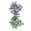 4ey4C  4ey5C 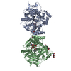 4ey6C  4ey7C  1b41S S: Starting model for refinement C: citing same article ( |
|---|---|
| Similar structure data |
-Links
 PDBj
PDBj


- Assembly
| Deposited unit | 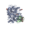
| ||||||||
|---|---|---|---|---|---|---|---|---|---|
| 1 |
| ||||||||
| Unit cell |
| ||||||||
| Details | dimer |
-Components
-Protein , 2 types, 2 molecules AB
| #1: Protein | / AChE Mass: 59447.105 Da / Num. of mol.: 1 / Fragment: UNP Residues 33-574 Source method: isolated from a genetically manipulated source Source: (gene. exp.) Homo sapiens (human) / Gene: ACHE / Production host: Homo sapiens (human) / Tissue (production host): HEK-293 / References: UniProt: P22303, acetylcholinesterase |
|---|---|
| #2: Protein | / Fas-2 / Fas2 / Acetylcholinesterase toxin F-VII / Fasciculin-II / FAS-II / Toxin TA1 Mass: 6768.769 Da / Num. of mol.: 1 / Source method: isolated from a natural source Source: (natural) Dendroaspis angusticeps (eastern green mamba)Secretion: VENOM / References: UniProt: P0C1Z0 |
-Sugars , 2 types, 2 molecules 
| #3: Polysaccharide | beta-D-mannopyranose-(1-4)-2-acetamido-2-deoxy-beta-D-glucopyranose-(1-4)-[alpha-L-fucopyranose-(1- ...beta-D-mannopyranose-(1-4)-2-acetamido-2-deoxy-beta-D-glucopyranose-(1-4)-[alpha-L-fucopyranose-(1-6)]2-acetamido-2-deoxy-beta-D-glucopyranose / Mass: 732.682 Da / Num. of mol.: 1 Source method: isolated from a genetically manipulated source |
|---|---|
| #5: Sugar | ChemComp-NAG / N-Acetylglucosamine Type: D-saccharide, beta linking / Mass: 221.208 Da / Num. of mol.: 1 Type: D-saccharide, beta linking / Mass: 221.208 Da / Num. of mol.: 1Source method: isolated from a genetically manipulated source Formula: C8H15NO6 |
-Non-polymers , 2 types, 48 molecules 

| #4: Chemical | ChemComp-SO4 / Sulfate Mass: 96.063 Da / Num. of mol.: 4 / Source method: obtained synthetically / Formula: SO4 Mass: 96.063 Da / Num. of mol.: 4 / Source method: obtained synthetically / Formula: SO4#6: Water | ChemComp-HOH / | WaterMass: 18.015 Da / Num. of mol.: 44 / Source method: isolated from a natural source / Formula: H2O |
|---|
-Experimental details
-Experiment
| Experiment | Method: X-RAY DIFFRACTION / Number of used crystals: 1 |
|---|
- Sample preparation
| Crystal | Density Matthews: 4.14 Å3/Da / Density % sol: 70.32 % |
|---|---|
| Crystal grow | Temperature: 283 K / Method: vapor diffusion, sitting drop Details: 1.6 to 2.0M ammonium sulphate, 0.1M HEPES pH 7.5 - 7.8, VAPOR DIFFUSION, SITTING DROP, temperature 283K PH range: 7.5 - 7.8 |
-Data collection
| Diffraction | Mean temperature: 100 K | |||||||||||||||||||||||||||||||||||||||||||||||||||||||||||||||||||||||||||||||||||||||||||||||||||||||||
|---|---|---|---|---|---|---|---|---|---|---|---|---|---|---|---|---|---|---|---|---|---|---|---|---|---|---|---|---|---|---|---|---|---|---|---|---|---|---|---|---|---|---|---|---|---|---|---|---|---|---|---|---|---|---|---|---|---|---|---|---|---|---|---|---|---|---|---|---|---|---|---|---|---|---|---|---|---|---|---|---|---|---|---|---|---|---|---|---|---|---|---|---|---|---|---|---|---|---|---|---|---|---|---|---|---|---|
| Diffraction source | Source: SYNCHROTRON / Site: NSLS  / Beamline: X4C / Wavelength: 0.99 Å / Beamline: X4C / Wavelength: 0.99 Å | |||||||||||||||||||||||||||||||||||||||||||||||||||||||||||||||||||||||||||||||||||||||||||||||||||||||||
| Detector | Type: MAR CCD 165 mm / Detector: CCD / Date: Jan 1, 2012 | |||||||||||||||||||||||||||||||||||||||||||||||||||||||||||||||||||||||||||||||||||||||||||||||||||||||||
| Radiation | Monochromator: Si(111) / Protocol: SINGLE WAVELENGTH / Monochromatic (M) / Laue (L): M / Scattering type: x-ray | |||||||||||||||||||||||||||||||||||||||||||||||||||||||||||||||||||||||||||||||||||||||||||||||||||||||||
| Radiation wavelength | Wavelength: 0.99 Å / Relative weight: 1 | |||||||||||||||||||||||||||||||||||||||||||||||||||||||||||||||||||||||||||||||||||||||||||||||||||||||||
| Reflection | Resolution: 2.5958→50 Å / Num. all: 34182 / Num. obs: 33704 / % possible obs: 98.6 % / Observed criterion σ(F): 0 / Observed criterion σ(I): -3 / Redundancy: 5 % / Rmerge(I) obs: 0.054 / Net I/σ(I): 11.1 | |||||||||||||||||||||||||||||||||||||||||||||||||||||||||||||||||||||||||||||||||||||||||||||||||||||||||
| Reflection shell |
|
-Phasing
| Phasing | Method: molecular replacement |
|---|
- Processing
| Software |
| |||||||||||||||||||||||||||||||||||||||||||||||||||||||||||||||||||||||||||||||||||||||||||
|---|---|---|---|---|---|---|---|---|---|---|---|---|---|---|---|---|---|---|---|---|---|---|---|---|---|---|---|---|---|---|---|---|---|---|---|---|---|---|---|---|---|---|---|---|---|---|---|---|---|---|---|---|---|---|---|---|---|---|---|---|---|---|---|---|---|---|---|---|---|---|---|---|---|---|---|---|---|---|---|---|---|---|---|---|---|---|---|---|---|---|---|---|
| Refinement | Method to determine structure: MOLECULAR REPLACEMENT Starting model: 1b41 Resolution: 2.5958→46.381 Å / Occupancy max: 1 / Occupancy min: 0.5 / SU ML: 0.35 / σ(F): 0 / Phase error: 30.82 / Stereochemistry target values: ML
| |||||||||||||||||||||||||||||||||||||||||||||||||||||||||||||||||||||||||||||||||||||||||||
| Solvent computation | Shrinkage radii: 1.1 Å / VDW probe radii: 1.3 Å / Solvent model: FLAT BULK SOLVENT MODEL | |||||||||||||||||||||||||||||||||||||||||||||||||||||||||||||||||||||||||||||||||||||||||||
| Displacement parameters |
| |||||||||||||||||||||||||||||||||||||||||||||||||||||||||||||||||||||||||||||||||||||||||||
| Refinement step | Cycle: LAST / Resolution: 2.5958→46.381 Å
| |||||||||||||||||||||||||||||||||||||||||||||||||||||||||||||||||||||||||||||||||||||||||||
| Refine LS restraints |
| |||||||||||||||||||||||||||||||||||||||||||||||||||||||||||||||||||||||||||||||||||||||||||
| LS refinement shell |
|
