 Movie
Movie Controller


+ Open data

- Basic information
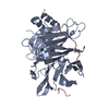
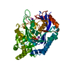
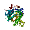
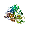
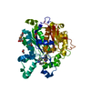
| Entry | Database: PDB / ID: 4exv | ||||||
|---|---|---|---|---|---|---|---|
| Title | Structure of Kluyveromyces lactis Hsv2p | ||||||
 Components Components | SVP1-like protein 2 | ||||||
 Keywords Keywords |  TRANSPORT PROTEIN / PROPPIN / WD-repeat / Phosphoinosides / Phosphatidylinositol / phosphate binding / Autophagy / Atg2 / Atg9 / Atg21 TRANSPORT PROTEIN / PROPPIN / WD-repeat / Phosphoinosides / Phosphatidylinositol / phosphate binding / Autophagy / Atg2 / Atg9 / Atg21 | ||||||
| Function / homology |  Function and homology information Function and homology informationvacuolar membrane / cytoplasmic vesicle membrane / autophagy / protein transportSimilarity search - Function | ||||||
| Biological species |  Kluyveromyces lactis (yeast) Kluyveromyces lactis (yeast) | ||||||
| Method | X-RAY DIFFRACTION / SYNCHROTRON / SAD / Resolution: 3 Å | ||||||
 Authors Authors | Baskaran, S. / Hurley, J.H. | ||||||
 Citation Citation | Journal: Mol.Cell / Year: 2012 Title: Two-Site Recognition of Phosphatidylinositol 3-Phosphate by PROPPINs in Autophagy. Authors: Baskaran, S. / Ragusa, M.J. / Boura, E. / Hurley, J.H. | ||||||
| History |
|
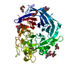
- Structure visualization
| Structure viewer | Molecule: MolmilJmol/JSmol |
|---|
- Downloads & links
-Download
| PDBx/mmCIF format | 4exv.cif.gz | 74.7 KB | Display | PDBx/mmCIF format |
|---|---|---|---|---|
| PDB format | pdb4exv.ent.gz | 56.4 KB | Display | PDB format |
| PDBx/mmJSON format | 4exv.json.gz | Tree view | PDBx/mmJSON format | |
| Others |  Other downloads Other downloads |
-Validation report
| Arichive directory | https://data.pdbj.org/pub/pdb/validation_reports/ex/4exvftp://data.pdbj.org/pub/pdb/validation_reports/ex/4exv | HTTPS FTP |
|---|
-Related structure data
| Similar structure data |
|---|


-Links
 PDBj
PDBj
- Assembly
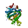
| Deposited unit | 
| ||||||||
|---|---|---|---|---|---|---|---|---|---|
| 1 |
| ||||||||
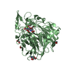
| Unit cell |
|
-Components
| #1: Protein | Mass: 39219.945 Da / Num. of mol.: 1 Source method: isolated from a genetically manipulated source Source: (gene. exp.) Kluyveromyces lactis (yeast)Strain: ATCC 8585 / CBS 2359 / DSM 70799 / NBRC 1267 / NRRL Y-1140 / WM37 Gene: HSV2, KLLA0E15972g / Plasmid: pGST2 / Production host:  Escherichia coli (E. coli) / Strain (production host): BL21-DE3 / References: UniProt: Q6CN23 Escherichia coli (E. coli) / Strain (production host): BL21-DE3 / References: UniProt: Q6CN23 |
|---|---|
| #2: Chemical | Sulfate  Mass: 96.063 Da / Num. of mol.: 3 / Source method: obtained synthetically / Formula: SO4 Mass: 96.063 Da / Num. of mol.: 3 / Source method: obtained synthetically / Formula: SO4 |
-Experimental details
-Experiment
| Experiment | Method: X-RAY DIFFRACTION / Number of used crystals: 1 |
|---|
- Sample preparation
| Crystal | Density Matthews: 4.13 Å3/Da / Density % sol: 70.22 % |
|---|---|
| Crystal grow | Temperature: 298 K / Method: vapor diffusion, hanging drop / pH: 6.2 Details: 50 mM MES pH 6.2, 1.8 M Magnesium sulfate, VAPOR DIFFUSION, HANGING DROP, temperature 298K |
-Data collection
| Diffraction | Mean temperature: 200 K |
|---|---|
| Diffraction source | Source: SYNCHROTRON / Site: APS  / Beamline: 22-ID / Wavelength: 1 Å / Beamline: 22-ID / Wavelength: 1 Å |
| Detector | Type: MARMOSAIC 300 mm CCD / Detector: CCD / Date: Oct 10, 2011 |
| Radiation | Monochromator: double crystal - liquid nitrogen cooled / Protocol: SINGLE WAVELENGTH / Monochromatic (M) / Laue (L): M / Scattering type: x-ray |
| Radiation wavelength | Wavelength: 1 Å / Relative weight: 1 |
| Reflection | Resolution: 3→50 Å / Num. obs: 13828 / % possible obs: 98 % / Observed criterion σ(F): 2 / Observed criterion σ(I): 1 / Biso Wilson estimate: 85.19 Å2 |
- Processing
| Software |
| ||||||||||||||||||||||||||||||||||||||||||
|---|---|---|---|---|---|---|---|---|---|---|---|---|---|---|---|---|---|---|---|---|---|---|---|---|---|---|---|---|---|---|---|---|---|---|---|---|---|---|---|---|---|---|---|
| Refinement | Method to determine structure: SAD / Resolution: 3→49.72 Å / Occupancy max: 1 / Occupancy min: 1 / SU ML: 0.69 / σ(F): 1.33 / Phase error: 25.67 / Stereochemistry target values: ML
| ||||||||||||||||||||||||||||||||||||||||||
| Solvent computation | Shrinkage radii: 0.83 Å / VDW probe radii: 1.1 Å / Solvent model: FLAT BULK SOLVENT MODEL / Bsol: 62.17 Å2 / ksol: 0.34 e/Å3 | ||||||||||||||||||||||||||||||||||||||||||
| Displacement parameters | Biso mean: 79.15 Å2
| ||||||||||||||||||||||||||||||||||||||||||
| Refinement step | Cycle: LAST / Resolution: 3→49.72 Å
| ||||||||||||||||||||||||||||||||||||||||||
| Refine LS restraints |
| ||||||||||||||||||||||||||||||||||||||||||
| LS refinement shell |
|
