 Movie
Movie Controller


+ Open data

- Basic information
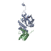
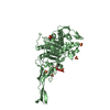
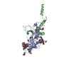
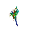
| Entry | Database: PDB / ID: 4eq6 | ||||||
|---|---|---|---|---|---|---|---|
| Title | The crystal structure of Psy3-Csm2 complex from budding yeast | ||||||
 Components Components |
| ||||||
 Keywords Keywords |  DNA BINDING PROTEIN DNA BINDING PROTEIN | ||||||
| Function / homology |  Function and homology information Function and homology informationpositive regulation of single-strand break repair via homologous recombination / Shu complex / error-free postreplication DNA repair / meiotic chromosome segregation / maintenance of rDNA / DNA recombinase assembly / recombinational repair / error-free translesion synthesis / site of double-strand break / nucleus ...positive regulation of single-strand break repair via homologous recombination / Shu complex / error-free postreplication DNA repair / meiotic chromosome segregation / maintenance of rDNA / DNA recombinase assembly / recombinational repair / error-free translesion synthesis / site of double-strand break / nucleus / cytosol / cytoplasmSimilarity search - Function | ||||||
| Biological species |  Saccharomyces cerevisiae (brewer's yeast) Saccharomyces cerevisiae (brewer's yeast) | ||||||
| Method | X-RAY DIFFRACTION / SYNCHROTRON / MOLECULAR REPLACEMENT / Resolution: 1.8 Å | ||||||
 Authors Authors | She, Z. / Gao, Z.Q. / Dong, Y.H. | ||||||
 Citation Citation | Journal: FEBS Lett. / Year: 2012 Title: Structural and SAXS analysis of the budding yeast SHU-complex proteins Authors: She, Z. / Gao, Z.Q. / Liu, Y. / Wang, W.J. / Liu, G.F. / Shtykova, E.V. / Xu, J.H. / Dong, Y.H. | ||||||
| History |
|
- Structure visualization
| Structure viewer | Molecule: MolmilJmol/JSmol |
|---|
- Downloads & links
-Download
| PDBx/mmCIF format | 4eq6.cif.gz | 180 KB | Display | PDBx/mmCIF format |
|---|---|---|---|---|
| PDB format | pdb4eq6.ent.gz | 141.4 KB | Display | PDB format |
| PDBx/mmJSON format | 4eq6.json.gz | Tree view | PDBx/mmJSON format | |
| Others |  Other downloads Other downloads |
-Validation report
| Arichive directory | https://data.pdbj.org/pub/pdb/validation_reports/eq/4eq6ftp://data.pdbj.org/pub/pdb/validation_reports/eq/4eq6 | HTTPS FTP |
|---|
-Related structure data
| Similar structure data |
|---|


-Links
 PDBj
PDBj- Assembly
| Deposited unit | 
| ||||||||
|---|---|---|---|---|---|---|---|---|---|
| 1 |
| ||||||||
| Unit cell |
| ||||||||
| Components on special symmetry positions |
|
-Components
| #1: Protein | Mass: 25114.875 Da / Num. of mol.: 1 Source method: isolated from a genetically manipulated source Source: (gene. exp.) Saccharomyces cerevisiae (brewer's yeast)Strain: ATCC 204508 / S288c / Gene: CSM2, YIL132C / Plasmid: pCDFDuet / Production host:  Escherichia coli (E. coli) / References: UniProt: P40465 Escherichia coli (E. coli) / References: UniProt: P40465 |
|---|---|
| #2: Protein | Mass: 29844.434 Da / Num. of mol.: 1 Source method: isolated from a genetically manipulated source Source: (gene. exp.) Saccharomyces cerevisiae (brewer's yeast)Strain: ATCC 204508 / S288c / Gene: PSY3, YLR376C, L8039.17 / Plasmid: pCDFDuet / Production host: Escherichia coli (E. coli) / References: UniProt: Q12318 |
| #3: Water | ChemComp-HOH / Water Mass: 18.015 Da / Num. of mol.: 162 / Source method: isolated from a natural source / Formula: H2O Mass: 18.015 Da / Num. of mol.: 162 / Source method: isolated from a natural source / Formula: H2O |
-Experimental details
-Experiment
| Experiment | Method: X-RAY DIFFRACTION / Number of used crystals: 1 |
|---|
- Sample preparation
| Crystal | Density Matthews: 2.36 Å3/Da / Density % sol: 47.98 % |
|---|---|
| Crystal grow | Temperature: 293 K / Method: vapor diffusion, sitting drop / pH: 8 Details: 100mM Tris-HCl, pH 8.0, 10% ethanol, 10% 2-methyl-2,4-pentadiol (MPD), VAPOR DIFFUSION, SITTING DROP, temperature 293K |
-Data collection
| Diffraction | Mean temperature: 100 K |
|---|---|
| Diffraction source | Source: SYNCHROTRON / Site: SSRF  / Beamline: BL17U / Wavelength: 0.9791 Å / Beamline: BL17U / Wavelength: 0.9791 Å |
| Detector | Type: ADSC QUANTUM 315r / Detector: CCD / Date: Mar 20, 2011 |
| Radiation | Monochromator: double crystal monochromator / Protocol: SINGLE WAVELENGTH / Monochromatic (M) / Laue (L): M / Scattering type: x-ray |
| Radiation wavelength | Wavelength: 0.9791 Å / Relative weight: 1 |
| Reflection | Resolution: 1.76→50 Å / Num. all: 46537 / Num. obs: 43595 / % possible obs: 94 % / Observed criterion σ(F): 0 / Observed criterion σ(I): 0 / Redundancy: 3.6 % / Biso Wilson estimate: 30.5 Å2 / Rmerge(I) obs: 0.043 / Net I/σ(I): 38.9 |
| Reflection shell | Resolution: 1.76→1.79 Å / Redundancy: 3.4 % / Rmerge(I) obs: 0.542 / Mean I/σ(I) obs: 2.5 / Num. unique all: 2353 / % possible all: 91.9 |
- Processing
| Software |
| ||||||||||||||||||||||||||||||||||||||||||||||||||||||||||||||||||||||||||||||||||||||||||||||||||||||||||||||||
|---|---|---|---|---|---|---|---|---|---|---|---|---|---|---|---|---|---|---|---|---|---|---|---|---|---|---|---|---|---|---|---|---|---|---|---|---|---|---|---|---|---|---|---|---|---|---|---|---|---|---|---|---|---|---|---|---|---|---|---|---|---|---|---|---|---|---|---|---|---|---|---|---|---|---|---|---|---|---|---|---|---|---|---|---|---|---|---|---|---|---|---|---|---|---|---|---|---|---|---|---|---|---|---|---|---|---|---|---|---|---|---|---|---|
| Refinement | Method to determine structure: MOLECULAR REPLACEMENT / Resolution: 1.8→29.855 Å / SU ML: 0.2 / σ(F): 1.35 / Phase error: 24.74 / Stereochemistry target values: ML
| ||||||||||||||||||||||||||||||||||||||||||||||||||||||||||||||||||||||||||||||||||||||||||||||||||||||||||||||||
| Solvent computation | Shrinkage radii: 0.98 Å / VDW probe radii: 1.2 Å / Solvent model: FLAT BULK SOLVENT MODEL / Bsol: 58.963 Å2 / ksol: 0.355 e/Å3 | ||||||||||||||||||||||||||||||||||||||||||||||||||||||||||||||||||||||||||||||||||||||||||||||||||||||||||||||||
| Displacement parameters |
| ||||||||||||||||||||||||||||||||||||||||||||||||||||||||||||||||||||||||||||||||||||||||||||||||||||||||||||||||
| Refinement step | Cycle: LAST / Resolution: 1.8→29.855 Å
| ||||||||||||||||||||||||||||||||||||||||||||||||||||||||||||||||||||||||||||||||||||||||||||||||||||||||||||||||
| Refine LS restraints |
| ||||||||||||||||||||||||||||||||||||||||||||||||||||||||||||||||||||||||||||||||||||||||||||||||||||||||||||||||
| LS refinement shell | Refine-ID: X-RAY DIFFRACTION / Total num. of bins used: 15
| ||||||||||||||||||||||||||||||||||||||||||||||||||||||||||||||||||||||||||||||||||||||||||||||||||||||||||||||||
| Refinement TLS params. | Method: refined / Origin x: -16.7614 Å / Origin y: 1.8044 Å / Origin z: -8.4787 Å
| ||||||||||||||||||||||||||||||||||||||||||||||||||||||||||||||||||||||||||||||||||||||||||||||||||||||||||||||||
| Refinement TLS group |
|
