- PDB-4eiu: Crystal structure of a DUF3823 family protein (BACUNI_03093) from... -
+
Open data
ID or keywords:
Loading...
-
Basic information
Entry
Database: PDB / ID: 4eiu
Title
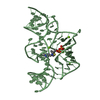
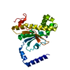
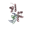
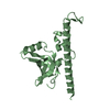
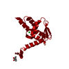
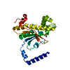
Crystal structure of a DUF3823 family protein (BACUNI_03093) from Bacteroides uniformis ATCC 8492 at 1.90 A resolution
Components
uncharacterized hypothetical protein
Keywords
STRUCTURAL GENOMICS / UNKNOWN FUNCTION / PF12866 family protein / DUF3823 / Joint Center for Structural Genomics / JCSG / Protein Structure Initiative / PSI-BIOLOGY
Function / homology
Function and homology information
Immunoglobulin-like - #2060 / DUF3823, N-terminal / Domain of unknown function DUF3823_C / Protein of unknown function (DUF3823) N-terminal domain / Domain of unknown function (DUF3823_C) / Carboxypeptidase-like, regulatory domain / Immunoglobulin-like / Sandwich / Mainly Beta Similarity search - Domain/homology
Mass: 18.015 Da / Num. of mol.: 142 / Source method: isolated from a natural source / Formula: H2O
Sequence details
THIS CONSTRUCT (RESIDUES 24-271) WAS EXPRESSED WITH A PURIFICATION TAG MGSDKIHHHHHHENLYFQG. THE TAG ...THIS CONSTRUCT (RESIDUES 24-271) WAS EXPRESSED WITH A PURIFICATION TAG MGSDKIHHHHHHENLYFQG. THE TAG WAS REMOVED WITH TEV PROTEASE LEAVING ONLY A GLYCINE (0) FOLLOWED BY THE TARGET SEQUENCE.
-
Experimental details
-
Experiment
Experiment
Method: X-RAY DIFFRACTION / Number of used crystals: 1
-
Sample preparation
Crystal
Density Matthews: 3.49 Å3/Da / Density % sol: 64.78 %
Crystal grow
Temperature: 277 K / Method: vapor diffusion, sitting drop / pH: 9.5 Details: 50.0% polyethylene glycol 200, 0.1M CHES pH 9.5, NANODROP, VAPOR DIFFUSION, SITTING DROP, temperature 277K
Monochromator: double crystal Si(111) / Protocol: SINGLE WAVELENGTH / Monochromatic (M) / Laue (L): M / Scattering type: x-ray
Radiation wavelength
Wavelength: 0.97915 Å / Relative weight: 1
Reflection
Resolution: 1.9→29.661 Å / Num. all: 32310 / Num. obs: 32310 / % possible obs: 99.9 % / Redundancy: 11.9 % / Biso Wilson estimate: 33.31 Å2 / Rsym value: 0.095 / Net I/σ(I): 12.4
Reflection shell
Diffraction-ID: 1
Resolution (Å)
Redundancy (%)
Rmerge(I) obs
Mean I/σ(I) obs
Num. measured all
Num. unique all
Rsym value
% possible all
1.9-1.95
11.9
0.972
2.6
27977
2347
0.972
100
1.95-2
12
0.742
1.1
27244
2279
0.742
100
2-2.06
12
0.556
1.4
26558
2216
0.556
100
2.06-2.12
12
0.452
1.7
25993
2172
0.452
100
2.12-2.19
12
0.359
2.1
25150
2095
0.359
100
2.19-2.27
12
0.295
2.5
24537
2046
0.295
100
2.27-2.36
12
0.24
3.1
23417
1950
0.24
100
2.36-2.45
12
0.203
3.6
22884
1905
0.203
100
2.45-2.56
12
0.18
4.1
21839
1826
0.18
100
2.56-2.69
12
0.15
4.5
20910
1747
0.15
100
2.69-2.83
11.9
0.132
5.1
19793
1665
0.132
100
2.83-3
11.9
0.113
5.7
18905
1585
0.113
100
3-3.21
11.9
0.098
6.3
17792
1495
0.098
100
3.21-3.47
11.9
0.079
7.8
16700
1403
0.079
100
3.47-3.8
11.8
0.068
9.4
15266
1294
0.068
100
3.8-4.25
11.7
0.061
10.7
13865
1185
0.061
100
4.25-4.91
11.6
0.069
9.3
12122
1046
0.069
99.9
4.91-6.01
11.4
0.074
8.7
10347
911
0.074
100
6.01-8.5
10.9
0.072
9.2
7954
728
0.072
100
8.5-29.661
9.3
0.064
10.4
3869
415
0.064
94.9
-
Phasing
Phasing
Method: SAD
-
Processing
Software
Name
Version
Classification
NB
MolProbity
3beta29
modelbuilding
PDB_EXTRACT
3.1
dataextraction
SHELX
phasing
SHARP
phasing
SCALA
3.3.20
datascaling
REFMAC
5.6.0117
refinement
MOSFLM
datareduction
SHELXD
phasing
autoSHARP
phasing
Refinement
Method to determine structure: SAD / Resolution: 1.9→29.661 Å / Cor.coef. Fo:Fc: 0.968 / Cor.coef. Fo:Fc free: 0.957 / Occupancy max: 1 / Occupancy min: 0.5 / SU B: 5.168 / SU ML: 0.08 / Cross valid method: THROUGHOUT / σ(F): 0 / ESU R: 0.109 / ESU R Free: 0.106 Stereochemistry target values: MAXIMUM LIKELIHOOD WITH PHASES Details: 1.HYDROGENS HAVE BEEN ADDED IN THE RIDING POSITIONS. 2.A MET-INHIBITION PROTOCOL WAS USED FOR SELENOMETHIONINE INCORPORATION DURING PROTEIN EXPRESSION. THE OCCUPANCY OF THE SE ATOMS IN THE ...Details: 1.HYDROGENS HAVE BEEN ADDED IN THE RIDING POSITIONS. 2.A MET-INHIBITION PROTOCOL WAS USED FOR SELENOMETHIONINE INCORPORATION DURING PROTEIN EXPRESSION. THE OCCUPANCY OF THE SE ATOMS IN THE MSE RESIDUES WAS REDUCED TO 0.75 FOR THE REDUCED SCATTERING POWER DUE TO PARTIAL S-MET INCORPORATION. 3.ATOM RECORD CONTAINS SUM OF TLS AND RESIDUAL B FACTORS. ANISOU RECORD CONTAINS SUM OF TLS AND RESIDUAL U FACTORS. 4.WATERS WERE EXCLUDED FROM AUTOMATIC TLS ASSIGNMENT. 5.PEG200 FRAGMENTS (PEG AND PGE) FROM THE CRYSTALLIZATION SOLUTION HAVE BEEN MODELED IN THE SOLVENT STRUCTURE.
Rfactor
Num. reflection
% reflection
Selection details
Rfree
0.2065
1634
5.1 %
RANDOM
Rwork
0.1789
-
-
-
obs
0.1803
32242
99.88 %
-
Solvent computation
Ion probe radii: 0.8 Å / Shrinkage radii: 0.8 Å / VDW probe radii: 1.2 Å / Solvent model: BABINET MODEL WITH MASK
In the structure databanks used in Yorodumi, some data are registered as the other names, "COVID-19 virus" and "2019-nCoV". Here are the details of the virus and the list of structure data.
Jan 31, 2019. EMDB accession codes are about to change! (news from PDBe EMDB page)
EMDB accession codes are about to change! (news from PDBe EMDB page)
The allocation of 4 digits for EMDB accession codes will soon come to an end. Whilst these codes will remain in use, new EMDB accession codes will include an additional digit and will expand incrementally as the available range of codes is exhausted. The current 4-digit format prefixed with “EMD-” (i.e. EMD-XXXX) will advance to a 5-digit format (i.e. EMD-XXXXX), and so on. It is currently estimated that the 4-digit codes will be depleted around Spring 2019, at which point the 5-digit format will come into force.
The EM Navigator/Yorodumi systems omit the EMD- prefix.
Related info.:Q: What is EMD? / ID/Accession-code notation in Yorodumi/EM Navigator
Yorodumi is a browser for structure data from EMDB, PDB, SASBDB, etc.
This page is also the successor to EM Navigator detail page, and also detail information page/front-end page for Omokage search.
The word "yorodu" (or yorozu) is an old Japanese word meaning "ten thousand". "mi" (miru) is to see.
Related info.:EMDB / PDB / SASBDB / Comparison of 3 databanks / Yorodumi Search / Aug 31, 2016. New EM Navigator & Yorodumi / Yorodumi Papers / Jmol/JSmol / Function and homology information / Changes in new EM Navigator and Yorodumi
 Movie
Movie Controller

 Yorodumi
 Open data
 Basic information
 Components
 Keywords
 STRUCTURAL GENOMICS / UNKNOWN FUNCTION / PF12866 family protein / DUF3823 / Joint Center for Structural Genomics / JCSG /
  Function and homology information
 Bacteroides uniformis (bacteria)
 Authors
 Citation
 Structure visualization
 Downloads & links
 Other downloads
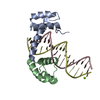


 PDBj
PDBj Assembly
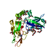

 Mass: 106.120 Da / Num. of mol.: 11 / Source method: obtained synthetically / Formula: C4H10O3
 Mass: 150.173 Da / Num. of mol.: 2 / Source method: obtained synthetically / Formula: C6H14O4
 Mass: 18.015 Da / Num. of mol.: 142 / Source method: isolated from a natural source / Formula: H2O
 Sample preparation
 / Beamline: BL14-1 / Wavelength: 0.97915
  Processing