- PDB-4ehs: Crystal structure of Helicobacter pylori DnaG Primase C terminal ... -
+
Open data
ID or keywords:
Loading...
-
Basic information
Entry
Database: PDB / ID: 4ehs
Title
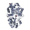
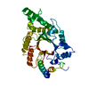
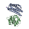
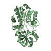
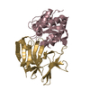
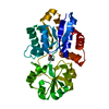
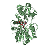
Crystal structure of Helicobacter pylori DnaG Primase C terminal domain
Components
DNA primasePrimase
Keywords
TRANSFERASE / primase / helicase binding domain
Function / homology
Function and homology information
primosome complex / DNA primase activity / DNA replication, synthesis of primer / Transferases; Transferring phosphorus-containing groups; Nucleotidyltransferases / DNA binding / zinc ion binding / cytoplasm Similarity search - Function
Method to determine structure: SAD / Resolution: 1.78→49.27 Å / Cor.coef. Fo:Fc: 0.955 / Cor.coef. Fo:Fc free: 0.927 / Occupancy max: 1 / Occupancy min: 1 / SU B: 2.919 / SU ML: 0.093 / Cross valid method: THROUGHOUT / ESU R: 0.139 / ESU R Free: 0.14 / Stereochemistry target values: MAXIMUM LIKELIHOOD / Details: HYDROGENS HAVE BEEN USED IF PRESENT IN THE INPUT
Rfactor
Num. reflection
% reflection
Selection details
Rfree
0.24929
1250
5.1 %
RANDOM
Rwork
0.19502
-
-
-
obs
0.19779
23200
99.59 %
-
all
-
24598
-
-
Solvent computation
Ion probe radii: 0.8 Å / Shrinkage radii: 0.8 Å / VDW probe radii: 1.2 Å / Solvent model: MASK
Displacement parameters
Biso mean: 23.766 Å2
Baniso -1
Baniso -2
Baniso -3
1-
-0.24 Å2
0 Å2
0 Å2
2-
-
1.06 Å2
0 Å2
3-
-
-
-0.81 Å2
Refine analyze
Free
Obs
Luzzati coordinate error
0.27 Å
0.2199 Å
Refinement step
Cycle: LAST / Resolution: 1.78→49.27 Å
Protein
Nucleic acid
Ligand
Solvent
Total
Num. atoms
1993
0
16
213
2222
Refine LS restraints
Refine-ID
Type
Dev ideal
Dev ideal target
Number
X-RAY DIFFRACTION
r_bond_refined_d
0.019
0.019
2039
X-RAY DIFFRACTION
r_bond_other_d
X-RAY DIFFRACTION
r_angle_refined_deg
2.184
2.053
2724
X-RAY DIFFRACTION
r_angle_other_deg
X-RAY DIFFRACTION
r_dihedral_angle_1_deg
5.484
5
244
X-RAY DIFFRACTION
r_dihedral_angle_2_deg
31.736
24.321
81
X-RAY DIFFRACTION
r_dihedral_angle_3_deg
15.638
15
417
X-RAY DIFFRACTION
r_dihedral_angle_4_deg
15.477
15
12
X-RAY DIFFRACTION
r_chiral_restr
0.126
0.2
314
X-RAY DIFFRACTION
r_gen_planes_refined
0.012
0.021
1420
X-RAY DIFFRACTION
r_gen_planes_other
X-RAY DIFFRACTION
r_nbd_refined
X-RAY DIFFRACTION
r_nbd_other
X-RAY DIFFRACTION
r_nbtor_refined
X-RAY DIFFRACTION
r_nbtor_other
X-RAY DIFFRACTION
r_xyhbond_nbd_refined
X-RAY DIFFRACTION
r_xyhbond_nbd_other
X-RAY DIFFRACTION
r_metal_ion_refined
X-RAY DIFFRACTION
r_metal_ion_other
X-RAY DIFFRACTION
r_symmetry_vdw_refined
X-RAY DIFFRACTION
r_symmetry_vdw_other
X-RAY DIFFRACTION
r_symmetry_hbond_refined
X-RAY DIFFRACTION
r_symmetry_hbond_other
X-RAY DIFFRACTION
r_symmetry_metal_ion_refined
X-RAY DIFFRACTION
r_symmetry_metal_ion_other
X-RAY DIFFRACTION
r_mcbond_it
1.467
1.5
1248
X-RAY DIFFRACTION
r_mcbond_other
X-RAY DIFFRACTION
r_mcangle_it
2.598
2
1998
X-RAY DIFFRACTION
r_scbond_it
4.318
3
812
X-RAY DIFFRACTION
r_scangle_it
6.855
4.5
753
X-RAY DIFFRACTION
r_rigid_bond_restr
X-RAY DIFFRACTION
r_sphericity_free
X-RAY DIFFRACTION
r_sphericity_bonded
LS refinement shell
Resolution: 1.778→1.824 Å / Total num. of bins used: 20
Rfactor
Num. reflection
% reflection
Rfree
0.35
80
-
Rwork
0.263
1469
-
obs
-
-
94.28 %
+
About Yorodumi
-
News
-
Feb 9, 2022. New format data for meta-information of EMDB entries
New format data for meta-information of EMDB entries
Version 3 of the EMDB header file is now the official format.
The previous official version 1.9 will be removed from the archive.
In the structure databanks used in Yorodumi, some data are registered as the other names, "COVID-19 virus" and "2019-nCoV". Here are the details of the virus and the list of structure data.
Jan 31, 2019. EMDB accession codes are about to change! (news from PDBe EMDB page)
EMDB accession codes are about to change! (news from PDBe EMDB page)
The allocation of 4 digits for EMDB accession codes will soon come to an end. Whilst these codes will remain in use, new EMDB accession codes will include an additional digit and will expand incrementally as the available range of codes is exhausted. The current 4-digit format prefixed with “EMD-” (i.e. EMD-XXXX) will advance to a 5-digit format (i.e. EMD-XXXXX), and so on. It is currently estimated that the 4-digit codes will be depleted around Spring 2019, at which point the 5-digit format will come into force.
The EM Navigator/Yorodumi systems omit the EMD- prefix.
Related info.:Q: What is EMD? / ID/Accession-code notation in Yorodumi/EM Navigator
Yorodumi is a browser for structure data from EMDB, PDB, SASBDB, etc.
This page is also the successor to EM Navigator detail page, and also detail information page/front-end page for Omokage search.
The word "yorodu" (or yorozu) is an old Japanese word meaning "ten thousand". "mi" (miru) is to see.
Related info.:EMDB / PDB / SASBDB / Comparison of 3 databanks / Yorodumi Search / Aug 31, 2016. New EM Navigator & Yorodumi / Yorodumi Papers / Jmol/JSmol / Function and homology information / Changes in new EM Navigator and Yorodumi
 Movie
Movie Controller

 Yorodumi
 Open data
 Basic information
 Components
 Primase
  Keywords
 Function and homology information

 Authors
 Citation
 Structure visualization
 Downloads & links
 Other downloads


 PDBj
PDBj


 Assembly

 Mass: 78.133 Da / Num. of mol.: 4 / Source method: obtained synthetically / Formula: C2H6OS
 Mass: 18.015 Da / Num. of mol.: 213 / Source method: isolated from a natural source / Formula: H2O
 Sample preparation
 / Beamline: BM14 / Wavelength: 0.9786 Å
 Processing