 Movie
Movie Controller

[English] 日本語
 Yorodumi
- PDB-4egl: Crystal structure of two tandem RNA recognition motifs of Human a... -
+ Open data

- Basic information
| Entry | Database: PDB / ID: 4egl | ||||||
|---|---|---|---|---|---|---|---|
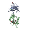
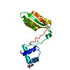
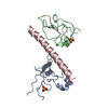
| Title | Crystal structure of two tandem RNA recognition motifs of Human antigen R | ||||||
 Components Components | ELAV-like protein 1 | ||||||
 Keywords Keywords | RNA BINDING PROTEIN / RRM / RNA Binding / Cytoplasm and Nucleus | ||||||
| Function / homology |  Function and homology information Function and homology informationHuR (ELAVL1) binds and stabilizes mRNA / negative regulation of miRNA-mediated gene silencing / post-transcriptional gene silencing / regulation of stem cell population maintenance / mRNA 3'-UTR AU-rich region binding / mRNA stabilization / lncRNA binding / miRNA binding / 3'-UTR-mediated mRNA stabilization / mRNA destabilization ...HuR (ELAVL1) binds and stabilizes mRNA / negative regulation of miRNA-mediated gene silencing / post-transcriptional gene silencing / regulation of stem cell population maintenance / mRNA 3'-UTR AU-rich region binding / mRNA stabilization / lncRNA binding / miRNA binding / 3'-UTR-mediated mRNA stabilization / mRNA destabilization / sarcoplasm / positive regulation of superoxide anion generation / positive regulation of translation / mRNA 3'-UTR binding / P-body / protein homooligomerization / cytoplasmic stress granule / protein import into nucleus / double-stranded RNA binding / cytoplasmic vesicle / postsynapse / ribonucleoprotein complex / mRNA binding / glutamatergic synapse / protein kinase binding / endoplasmic reticulum / protein homodimerization activity / RNA binding / nucleoplasm / membrane / nucleus / cytosol / cytoplasmSimilarity search - Function | ||||||
| Biological species |  Homo sapiens (human) Homo sapiens (human) | ||||||
| Method | X-RAY DIFFRACTION / MOLECULAR REPLACEMENT / Resolution: 2.9 Å | ||||||
 Authors Authors | Wang, H. / Zeng, F. / Liu, H. / Teng, M. / Li, X. | ||||||
 Citation Citation | Journal: To be Published Title: Crystal structure of two tandem RNA recognition motifs of Human antigen R Authors: Wang, H. / Zeng, F. / Liu, H. / Teng, M. / Li, X. | ||||||
| History |
|
- Structure visualization
| Structure viewer | Molecule: MolmilJmol/JSmol |
|---|
- Downloads & links
-Download
| PDBx/mmCIF format | 4egl.cif.gz | 75.4 KB | Display | PDBx/mmCIF format |
|---|---|---|---|---|
| PDB format | pdb4egl.ent.gz | 55.1 KB | Display | PDB format |
| PDBx/mmJSON format | 4egl.json.gz | Tree view | PDBx/mmJSON format | |
| Others |  Other downloads Other downloads |
-Validation report
| Arichive directory | https://data.pdbj.org/pub/pdb/validation_reports/eg/4eglftp://data.pdbj.org/pub/pdb/validation_reports/eg/4egl | HTTPS FTP |
|---|
-Related structure data
| Related structure data | 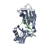 1fxlS S: Starting model for refinement |
|---|---|
| Similar structure data |


-Links
 PDBj
PDBj
- Assembly
| Deposited unit | 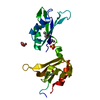
| ||||||||
|---|---|---|---|---|---|---|---|---|---|
| 1 |
| ||||||||
| Unit cell |
|
-Components
| #1: Protein | / Hu-antigen R / HuR Mass: 19839.420 Da / Num. of mol.: 1 Fragment: N-terminal RRM1 and RRM2 domain, UNP residues 18-186 Source method: isolated from a genetically manipulated source Source: (gene. exp.) Homo sapiens (human) / Gene: ELAVL1, HUR / Production host:  Escherichia coli (E. coli) / References: UniProt: Q15717 Escherichia coli (E. coli) / References: UniProt: Q15717 |
|---|---|
| #2: Chemical | ChemComp-SO4 / Sulfate  Mass: 96.063 Da / Num. of mol.: 1 / Source method: obtained synthetically / Formula: SO4 Mass: 96.063 Da / Num. of mol.: 1 / Source method: obtained synthetically / Formula: SO4 |
| #3: Chemical | Glycerol  Mass: 92.094 Da / Num. of mol.: 3 / Source method: obtained synthetically / Formula: C3H8O3 Mass: 92.094 Da / Num. of mol.: 3 / Source method: obtained synthetically / Formula: C3H8O3 |
-Experimental details
-Experiment
| Experiment | Method: X-RAY DIFFRACTION / Number of used crystals: 1 |
|---|
- Sample preparation
| Crystal | Density Matthews: 2.14 Å3/Da / Density % sol: 42.58 % |
|---|---|
| Crystal grow | Temperature: 287 K / Method: vapor diffusion, hanging drop / pH: 8.5 Details: 1.5M Lithium sulfate monohydrate, 0.1M Tris, pH 8.5, VAPOR DIFFUSION, HANGING DROP, temperature 287K |
-Data collection
| Diffraction | Mean temperature: 100 K |
|---|---|
| Diffraction source | Source: ROTATING ANODE / Type: RIGAKU RUH3R / Wavelength: 0.99985 Å |
| Detector | Type: MAR scanner 345 mm plate / Detector: IMAGE PLATE / Date: Apr 21, 2010 |
| Radiation | Monochromator: Ni FILTER / Protocol: SINGLE WAVELENGTH / Monochromatic (M) / Laue (L): M / Scattering type: x-ray |
| Radiation wavelength | Wavelength: 0.99985 Å / Relative weight: 1 |
| Reflection | Resolution: 2.9→50 Å / Num. all: 4179 / Num. obs: 4062 / % possible obs: 97.3 % |
| Reflection shell | Resolution: 2.9→3 Å / Redundancy: 3.4 % / Rmerge(I) obs: 0.465 / Mean I/σ(I) obs: 2.26 / Rsym value: 0.366 / % possible all: 92.9 |
- Processing
| Software |
| ||||||||||||||||||||||||||||||||||||||||||||||||||||||||||||||||||||||||||||||||||||||||||||||||||||||||||||||||||||||||||||||||||||||||||||||||||||||||||||||||||||||||||
|---|---|---|---|---|---|---|---|---|---|---|---|---|---|---|---|---|---|---|---|---|---|---|---|---|---|---|---|---|---|---|---|---|---|---|---|---|---|---|---|---|---|---|---|---|---|---|---|---|---|---|---|---|---|---|---|---|---|---|---|---|---|---|---|---|---|---|---|---|---|---|---|---|---|---|---|---|---|---|---|---|---|---|---|---|---|---|---|---|---|---|---|---|---|---|---|---|---|---|---|---|---|---|---|---|---|---|---|---|---|---|---|---|---|---|---|---|---|---|---|---|---|---|---|---|---|---|---|---|---|---|---|---|---|---|---|---|---|---|---|---|---|---|---|---|---|---|---|---|---|---|---|---|---|---|---|---|---|---|---|---|---|---|---|---|---|---|---|---|---|---|---|
| Refinement | Method to determine structure: MOLECULAR REPLACEMENT Starting model: PDB ENTRY 1FXL Resolution: 2.9→39.33 Å / Cor.coef. Fo:Fc: 0.903 / Cor.coef. Fo:Fc free: 0.854 / Occupancy max: 1 / Occupancy min: 0.22 / SU B: 52.296 / SU ML: 0.448 / Cross valid method: THROUGHOUT / ESU R Free: 0.486 / Stereochemistry target values: MAXIMUM LIKELIHOOD / Details: HYDROGENS HAVE BEEN ADDED IN THE RIDING POSITIONS
| ||||||||||||||||||||||||||||||||||||||||||||||||||||||||||||||||||||||||||||||||||||||||||||||||||||||||||||||||||||||||||||||||||||||||||||||||||||||||||||||||||||||||||
| Solvent computation | Ion probe radii: 0.8 Å / Shrinkage radii: 0.8 Å / VDW probe radii: 1.4 Å / Solvent model: MASK | ||||||||||||||||||||||||||||||||||||||||||||||||||||||||||||||||||||||||||||||||||||||||||||||||||||||||||||||||||||||||||||||||||||||||||||||||||||||||||||||||||||||||||
| Displacement parameters | Biso mean: 59.645 Å2
| ||||||||||||||||||||||||||||||||||||||||||||||||||||||||||||||||||||||||||||||||||||||||||||||||||||||||||||||||||||||||||||||||||||||||||||||||||||||||||||||||||||||||||
| Refinement step | Cycle: LAST / Resolution: 2.9→39.33 Å
| ||||||||||||||||||||||||||||||||||||||||||||||||||||||||||||||||||||||||||||||||||||||||||||||||||||||||||||||||||||||||||||||||||||||||||||||||||||||||||||||||||||||||||
| Refine LS restraints |
| ||||||||||||||||||||||||||||||||||||||||||||||||||||||||||||||||||||||||||||||||||||||||||||||||||||||||||||||||||||||||||||||||||||||||||||||||||||||||||||||||||||||||||
| LS refinement shell | Resolution: 2.896→2.971 Å / Total num. of bins used: 20
| ||||||||||||||||||||||||||||||||||||||||||||||||||||||||||||||||||||||||||||||||||||||||||||||||||||||||||||||||||||||||||||||||||||||||||||||||||||||||||||||||||||||||||
| Refinement TLS params. | Method: refined / Origin x: -4.8111 Å / Origin y: 15.3323 Å / Origin z: -2.3802 Å
|
