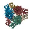
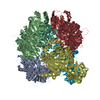
[English] 日本語
Yorodumi
- PDB-4efd: Crystal Structure of an M17 aminopeptidase from Trypanosoma Bruce... -

+
Open data


ID or keywords:

Loading...

-
Basic information

Entry
Database: PDB / ID: 4efd
TitleCrystal Structure of an M17 aminopeptidase from Trypanosoma Brucei, Tb427tmp.02.4440
ComponentsAminopeptidase
KeywordsHYDROLASE / Structural Genomics / Structural Genomics Consortium / SGC / aminopeptidase
Function / homology
Function and homology information


metalloaminopeptidase activity / aminopeptidase activity / manganese ion binding / peptidase activity / proteolysis / cytoplasm
Similarity search - Function
Zn-dependent exopeptidases / Probable aminopeptidase NPEPL1, N-terminal / M17 aminopeptidase N-terminal domain 2 / Cytosol aminopeptidase signature. / Peptidase M17, leucyl aminopeptidase, C-terminal / Peptidase M17, leucine aminopeptidase/peptidase B / Cytosol aminopeptidase family, catalytic domain / Zn peptidases / Aminopeptidase / Rossmann fold ...Zn-dependent exopeptidases / Probable aminopeptidase NPEPL1, N-terminal / M17 aminopeptidase N-terminal domain 2 / Cytosol aminopeptidase signature. / Peptidase M17, leucyl aminopeptidase, C-terminal / Peptidase M17, leucine aminopeptidase/peptidase B / Cytosol aminopeptidase family, catalytic domain / Zn peptidases / Aminopeptidase / Rossmann fold / 3-Layer(aba) Sandwich / Alpha Beta
Similarity search - Domain/homology
: / Aminopeptidase, putative
Similarity search - Component
Biological speciesTrypanosoma brucei brucei (eukaryote)
MethodX-RAY DIFFRACTION / SYNCHROTRON / MOLECULAR REPLACEMENT / molecular replacement / Resolution: 2.45 Å
AuthorsWernimont, A.K. / Osman, K.T. / Loppnau, P. / Arrowsmith, C.H. / Edwards, A.M. / Bountra, C. / Hui, R. / Lin, Y.H. / Structural Genomics Consortium (SGC)
CitationJournal: To be Published
Title: Crystal Structure of an M17 aminopeptidase from Trypanosoma Brucei, Tb427tmp.02.4440
Authors: Wernimont, A.K. / Osman, K.T. / Loppnau, P. / Arrowsmith, C.H. / Edwards, A.M. / Bountra, C. / Hui, R. / Lin, Y.H.
History
DepositionMar 29, 2012Deposition site: RCSB / Processing site: RCSB
Revision 1.0May 30, 2012Provider: repository / Type: Initial release
Revision 1.1Nov 15, 2017Group: Refinement description / Category: software
Item: _software.classification / _software.contact_author ..._software.classification / _software.contact_author / _software.contact_author_email / _software.date / _software.language / _software.location / _software.name / _software.type / _software.version
Revision 1.2Feb 28, 2024Group: Data collection / Database references / Derived calculations
Category: chem_comp_atom / chem_comp_bond ...chem_comp_atom / chem_comp_bond / database_2 / struct_site
Item: _database_2.pdbx_DOI / _database_2.pdbx_database_accession ..._database_2.pdbx_DOI / _database_2.pdbx_database_accession / _struct_site.pdbx_auth_asym_id / _struct_site.pdbx_auth_comp_id / _struct_site.pdbx_auth_seq_id

-
Structure visualization

Structure viewerMolecule:
MolmilJmol/JSmol

Downloads & links

-
Assembly

Deposited unit
A: Aminopeptidase
B: Aminopeptidase
C: Aminopeptidase
D: Aminopeptidase
E: Aminopeptidase
F: Aminopeptidase
hetero molecules


Theoretical massNumber of molelcules
Total (without water)335,22642
Polymers332,8516
Non-polymers2,37536
Water13,854769
1


  • Idetical with deposited unit
  • defined by author&software
TypeNameSymmetry operationNumber
identity operation1_555x,y,z1
Buried area31000 Å2
ΔGint-306 kcal/mol
Surface area94050 Å2
MethodPISA
Unit cell
Length a, b, c (Å)161.916, 161.740, 176.315
Angle α, β, γ (deg.)90.00, 90.00, 90.00
Int Tables number19
Space group name H-MP212121
Noncrystallographic symmetry (NCS)NCS domain:
IDEns-IDDetails
11A
21B
31C
41D
51E
61F

NCS domain segments:
Dom-IDComponent-IDEns-IDRefine codeAuth asym-IDAuth seq-ID
1113A349 - 355
2113B349 - 355
3113C349 - 355
4113D349 - 355
5113E349 - 355
6113F349 - 355
1213A495 - 501
2213B495 - 501
3213C495 - 501
4213D495 - 501
5213E495 - 501
6213F495 - 501
1314A289 - 305
2314B289 - 305
3314C289 - 305
4314D289 - 305
5314E289 - 305
6314F289 - 305

-
Components

-
Protein , 1 types, 6 molecules ABCDEF

#1: Protein
Aminopeptidase /


Mass: 55475.191 Da / Num. of mol.: 6
Source method: isolated from a genetically manipulated source
Source: (gene. exp.) Trypanosoma brucei brucei (eukaryote) / Strain: 927/4 GUTat10.1 / Gene: Tb11.02.4440 / Production host: Escherichia coli (E. coli) / References: UniProt: Q385B0*PLUS

-
Non-polymers , 6 types, 805 molecules

#2: Chemical
ChemComp-SO4 / SULFATE ION / Sulfate


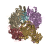
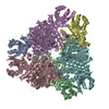
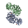
Mass: 96.063 Da / Num. of mol.: 12 / Source method: obtained synthetically / Formula: SO4
#3: Chemical
ChemComp-MN / MANGANESE (II) ION


Mass: 54.938 Da / Num. of mol.: 8 / Source method: obtained synthetically / Formula: Mn
#4: Chemical
ChemComp-GOL / GLYCEROL / GLYCERIN / PROPANE-1,2,3-TRIOL / Glycerol


Mass: 92.094 Da / Num. of mol.: 8 / Source method: obtained synthetically / Formula: C3H8O3
#5: Chemical
ChemComp-UNX / UNKNOWN ATOM OR ION


Num. of mol.: 6 / Source method: obtained synthetically
#6: Chemical ChemComp-NA / SODIUM ION


Mass: 22.990 Da / Num. of mol.: 2 / Source method: obtained synthetically / Formula: Na
#7: Water ChemComp-HOH / water / Water


Mass: 18.015 Da / Num. of mol.: 769 / Source method: isolated from a natural source / Formula: H2O

-
Experimental details

-
Experiment

ExperimentMethod: X-RAY DIFFRACTION / Number of used crystals: 1

-
Sample preparation

CrystalDensity Matthews: 3.47 Å3/Da / Density % sol: 64.53 %
Crystal growTemperature: 293 K / pH: 5.6
Details: 100 mM Na Citrate pH 5.6, 0.2 M Ammonium Sulfate, 2.0 M K/Na Tartrate, 2 mM MnCl2, 5 mM MgCl2, VAPOR DIFFUSION, SITTING DROP, temperature 293K

-
Data collection

DiffractionMean temperature: 100 K
Diffraction sourceSource: SYNCHROTRON / Site: APS / Beamline: 19-ID / Wavelength: 0.97931
DetectorType: ADSC QUANTUM 315 / Detector: CCD / Date: Feb 24, 2012
RadiationProtocol: SINGLE WAVELENGTH / Monochromatic (M) / Laue (L): M / Scattering type: x-ray
Radiation wavelengthWavelength: 0.97931 Å / Relative weight: 1
ReflectionResolution: 2.45→50 Å / Num. obs: 169603 / % possible obs: 99.5 % / Observed criterion σ(I): 0 / Redundancy: 7.9 % / Rmerge(I) obs: 0.109 / Net I/σ(I): 6
Reflection shellResolution: 2.45→2.49 Å / Redundancy: 7.4 % / Rmerge(I) obs: 0.975 / Mean I/σ(I) obs: 2 / % possible all: 98.5

-
Phasing

PhasingMethod: molecular replacement

-
Processing

Software
NameVersionClassificationNB
DENZOdata reduction
SCALEPACKdata scaling
REFMACrefinement
PDB_EXTRACT3.1data extraction
BALBESphasing
RefinementMethod to determine structure: MOLECULAR REPLACEMENT / Resolution: 2.45→30 Å / Cor.coef. Fo:Fc: 0.935 / Cor.coef. Fo:Fc free: 0.916 / Occupancy max: 1 / Occupancy min: 0.3 / SU B: 8.624 / SU ML: 0.195 / SU R Cruickshank DPI: 0.3435 / Cross valid method: THROUGHOUT / σ(F): 0 / ESU R: 0.343 / ESU R Free: 0.243 / Stereochemistry target values: MAXIMUM LIKELIHOOD / Details: HYDROGENS HAVE BEEN ADDED IN THE RIDING
RfactorNum. reflection% reflectionSelection details
Rfree0.253 8455 5 %RANDOM
Rwork0.227 ---
obs0.228 168491 99.3 %-
all-169661 --
Solvent computationIon probe radii: 0.8 Å / Shrinkage radii: 0.8 Å / VDW probe radii: 1.4 Å / Solvent model: MASK
Displacement parametersBiso mean: 44.97 Å2
Baniso -1Baniso -2Baniso -3
1--0.86 Å20 Å20 Å2
2---1.8 Å20 Å2
3---2.66 Å2
Refinement stepCycle: LAST / Resolution: 2.45→30 Å
ProteinNucleic acidLigandSolventTotal
Num. atoms22664 0 124 769 23557
Refine LS restraints
Refine-IDTypeDev idealDev ideal targetNumber
X-RAY DIFFRACTIONr_bond_refined_d0.0050.02223461
X-RAY DIFFRACTIONr_bond_other_d0.0020.0215067
X-RAY DIFFRACTIONr_angle_refined_deg0.7911.96832036
X-RAY DIFFRACTIONr_angle_other_deg0.796336966
X-RAY DIFFRACTIONr_dihedral_angle_1_deg4.40953138
X-RAY DIFFRACTIONr_dihedral_angle_2_deg34.16724.364935
X-RAY DIFFRACTIONr_dihedral_angle_3_deg12.289153450
X-RAY DIFFRACTIONr_dihedral_angle_4_deg13.08315117
X-RAY DIFFRACTIONr_chiral_restr0.0550.23738
X-RAY DIFFRACTIONr_gen_planes_refined0.0030.02126832
X-RAY DIFFRACTIONr_gen_planes_other0.0010.024585
X-RAY DIFFRACTIONr_nbd_refined
X-RAY DIFFRACTIONr_nbd_other
X-RAY DIFFRACTIONr_nbtor_refined
X-RAY DIFFRACTIONr_nbtor_other
X-RAY DIFFRACTIONr_xyhbond_nbd_refined
X-RAY DIFFRACTIONr_xyhbond_nbd_other
X-RAY DIFFRACTIONr_metal_ion_refined
X-RAY DIFFRACTIONr_metal_ion_other
X-RAY DIFFRACTIONr_symmetry_vdw_refined
X-RAY DIFFRACTIONr_symmetry_vdw_other
X-RAY DIFFRACTIONr_symmetry_hbond_refined
X-RAY DIFFRACTIONr_symmetry_hbond_other
X-RAY DIFFRACTIONr_symmetry_metal_ion_refined
X-RAY DIFFRACTIONr_symmetry_metal_ion_other
X-RAY DIFFRACTIONr_mcbond_it0.2281.515506
X-RAY DIFFRACTIONr_mcbond_other0.0291.56329
X-RAY DIFFRACTIONr_mcangle_it0.419224838
X-RAY DIFFRACTIONr_scbond_it0.31937955
X-RAY DIFFRACTIONr_scangle_it0.584.57186
X-RAY DIFFRACTIONr_rigid_bond_restr
X-RAY DIFFRACTIONr_sphericity_free
X-RAY DIFFRACTIONr_sphericity_bonded
Refine LS restraints NCS

Ens-ID: 1 / Refine-ID: X-RAY DIFFRACTION

Dom-IDAuth asym-IDNumberTypeRms dev position (Å)Weight position
1A69tight positional0.010.05
2B69tight positional0.010.05
3C69tight positional0.010.05
4D69tight positional0.010.05
5E69tight positional0.010.05
6F69tight positional0.010.05
1A189medium positional0.5
2B189medium positional0.5
3C189medium positional0.5
4D189medium positional0.5
5E189medium positional0.5
6F189medium positional0.5
1A69loose positional0.125
2B69loose positional0.125
3C69loose positional0.125
4D69loose positional0.125
5E69loose positional0.125
6F69loose positional0.125
1A69tight thermal0.030.5
2B69tight thermal0.020.5
3C69tight thermal0.030.5
4D69tight thermal0.040.5
5E69tight thermal0.040.5
6F69tight thermal0.030.5
1A189medium thermal0.012
2B189medium thermal0.012
3C189medium thermal0.012
4D189medium thermal0.022
5E189medium thermal0.012
6F189medium thermal0.022
1A69loose thermal0.0110
2B69loose thermal0.0110
3C69loose thermal0.0110
4D69loose thermal0.0110
5E69loose thermal0.0110
6F69loose thermal0.0110
LS refinement shellResolution: 2.45→2.51 Å / Total num. of bins used: 20
RfactorNum. reflection% reflection
Rfree0.339 585 -
Rwork0.329 11611 -
obs--98.66 %

+
About Yorodumi

-
News

-
Feb 9, 2022. New format data for meta-information of EMDB entries

New format data for meta-information of EMDB entries

  • Version 3 of the EMDB header file is now the official format.
  • The previous official version 1.9 will be removed from the archive.

Related info.:EMDB header

External links:wwPDB to switch to version 3 of the EMDB data model

-
Aug 12, 2020. Covid-19 info

Covid-19 info

URL: https://pdbj.org/emnavi/covid19.php

New page: Covid-19 featured information page in EM Navigator.

Related info.:Covid-19 info / Mar 5, 2020. Novel coronavirus structure data

+
Mar 5, 2020. Novel coronavirus structure data

Novel coronavirus structure data

Related info.:Yorodumi Speices / Aug 12, 2020. Covid-19 info

External links:COVID-19 featured content - PDBj / Molecule of the Month (242):Coronavirus Proteases

+
Jan 31, 2019. EMDB accession codes are about to change! (news from PDBe EMDB page)

EMDB accession codes are about to change! (news from PDBe EMDB page)

  • The allocation of 4 digits for EMDB accession codes will soon come to an end. Whilst these codes will remain in use, new EMDB accession codes will include an additional digit and will expand incrementally as the available range of codes is exhausted. The current 4-digit format prefixed with “EMD-” (i.e. EMD-XXXX) will advance to a 5-digit format (i.e. EMD-XXXXX), and so on. It is currently estimated that the 4-digit codes will be depleted around Spring 2019, at which point the 5-digit format will come into force.
  • The EM Navigator/Yorodumi systems omit the EMD- prefix.

Related info.:Q: What is EMD? / ID/Accession-code notation in Yorodumi/EM Navigator

External links:EMDB Accession Codes are Changing Soon! / Contact to PDBj

+
Jul 12, 2017. Major update of PDB

Major update of PDB

  • wwPDB released updated PDB data conforming to the new PDBx/mmCIF dictionary.
  • This is a major update changing the version number from 4 to 5, and with Remediation, in which all the entries are updated.
  • In this update, many items about electron microscopy experimental information are reorganized (e.g. em_software).
  • Now, EM Navigator and Yorodumi are based on the updated data.

External links:wwPDB Remediation / Enriched Model Files Conforming to OneDep Data Standards Now Available in the PDB FTP Archive

-
Yorodumi

Thousand views of thousand structures

  • Yorodumi is a browser for structure data from EMDB, PDB, SASBDB, etc.
  • This page is also the successor to EM Navigator detail page, and also detail information page/front-end page for Omokage search.
  • The word "yorodu" (or yorozu) is an old Japanese word meaning "ten thousand". "mi" (miru) is to see.

Related info.:EMDB / PDB / SASBDB / Comparison of 3 databanks / Yorodumi Search / Aug 31, 2016. New EM Navigator & Yorodumi / Yorodumi Papers / Jmol/JSmol / Function and homology information / Changes in new EM Navigator and Yorodumi

Read more