 Movie
Movie Controller

[English] 日本語
 Yorodumi
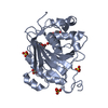
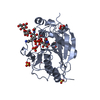
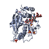
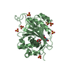
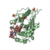
- PDB-4eea: Crystal structure of human M340H-beta-1,4-galactosyltransferase-1... -
+ Open data

- Basic information
| Entry | Database: PDB / ID: 4eea | |||||||||
|---|---|---|---|---|---|---|---|---|---|---|
| Title | Crystal structure of human M340H-beta-1,4-galactosyltransferase-1 (M340H-B4GAL-T1) in complex with GLCNAC-BETA1,6-Gal-Beta1,4-Glc-BETA | |||||||||
 Components Components | Beta-1,4-galactosyltransferase 1 | |||||||||
 Keywords Keywords |  TRANSFERASE / enzyme-carbohydrate complex / GT-A fold / glycosyltransferase / UDP-Galactose TRANSFERASE / enzyme-carbohydrate complex / GT-A fold / glycosyltransferase / UDP-Galactose | |||||||||
| Function / homology |  Function and homology information Function and homology informationDefective B4GALT1 causes CDG-2d / galactosyltransferase activity / Interaction With Cumulus Cells And The Zona Pellucida / Defective B4GALT1 causes B4GALT1-CDG (CDG-2d) / regulation of acrosome reaction / penetration of zona pellucida / Lactose synthesis / Keratan sulfate biosynthesis / lactose synthase / neolactotriaosylceramide beta-1,4-galactosyltransferase ...Defective B4GALT1 causes CDG-2d / galactosyltransferase activity / Interaction With Cumulus Cells And The Zona Pellucida / Defective B4GALT1 causes B4GALT1-CDG (CDG-2d) / regulation of acrosome reaction / penetration of zona pellucida / Lactose synthesis / Keratan sulfate biosynthesis / lactose synthase / neolactotriaosylceramide beta-1,4-galactosyltransferase / beta-N-acetylglucosaminylglycopeptide beta-1,4-galactosyltransferase / N-acetyllactosamine synthase / N-acetyllactosamine synthase activity / positive regulation of circulating fibrinogen levels / beta-N-acetylglucosaminylglycopeptide beta-1,4-galactosyltransferase activity / N-Glycan antennae elongation / UDP-galactosyltransferase activity / Golgi trans cisterna / macrophage migration / lactose synthase activity / development of secondary sexual characteristics / lactose biosynthetic process / oligosaccharide biosynthetic process / desmosome / galactose metabolic process / acute inflammatory response / positive regulation of epithelial cell proliferation involved in wound healing / Pre-NOTCH Processing in Golgi / binding of sperm to zona pellucida / angiogenesis involved in wound healing / protein N-linked glycosylation / Transferases; Glycosyltransferases; Hexosyltransferases / azurophil granule membrane / Golgi cisterna membrane / beta-tubulin binding / epithelial cell development / alpha-tubulin binding / cytoskeletal protein binding / extracellular matrix organization / secretory granule membrane / epithelial cell proliferation / filopodium / brush border membrane / lipid metabolic process / negative regulation of epithelial cell proliferation / manganese ion binding / basolateral plasma membrane / cell adhesion / positive regulation of apoptotic process / external side of plasma membrane / Golgi membrane / Neutrophil degranulation / Golgi apparatus / protein-containing complex / extracellular space / extracellular exosome / membrane / identical protein binding / plasma membraneSimilarity search - Function | |||||||||
| Biological species |  Homo sapiens (human) Homo sapiens (human) | |||||||||
| Method | X-RAY DIFFRACTION / Molecular Placement / Resolution: 2 Å | |||||||||
 Authors Authors | Ramakrishnan, B. / Qasba, P.K. | |||||||||
 Citation Citation | Journal: J.Biol.Chem. / Year: 2012 Title: Binding of N-acetylglucosamine (GlcNAc) beta 1-6-branched oligosaccharide acceptors to beta 4-galactosyltransferase I reveals a new ligand binding mode. Authors: Ramakrishnan, B. / Boeggeman, E. / Qasba, P.K. | |||||||||
| History |
|
- Structure visualization
| Structure viewer | Molecule: MolmilJmol/JSmol |
|---|
- Downloads & links
-Download
| PDBx/mmCIF format | 4eea.cif.gz | 204.5 KB | Display | PDBx/mmCIF format |
|---|---|---|---|---|
| PDB format | pdb4eea.ent.gz | 161.9 KB | Display | PDB format |
| PDBx/mmJSON format | 4eea.json.gz | Tree view | PDBx/mmJSON format | |
| Others |  Other downloads Other downloads |
-Validation report
| Arichive directory | https://data.pdbj.org/pub/pdb/validation_reports/ee/4eeaftp://data.pdbj.org/pub/pdb/validation_reports/ee/4eea | HTTPS FTP |
|---|
-Related structure data
| Related structure data |  4ee3C  4ee4C  4ee5C  4eegC  4eemC  4eeoC  2aecS C: citing same article ( S: Starting model for refinement |
|---|---|
| Similar structure data |


-Links
 PDBj
PDBj


- Assembly
| Deposited unit | 
| ||||||||
|---|---|---|---|---|---|---|---|---|---|
| 1 | 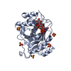
| ||||||||
| 2 | 
| ||||||||
| 3 | 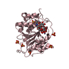
| ||||||||
| 4 | 
| ||||||||
| 5 |
| ||||||||
| Unit cell |
|
-Components
-Protein / Sugars , 2 types, 6 molecules ABC
| #1: Protein | Mass: 32773.230 Da / Num. of mol.: 3 / Fragment: catalytic domain / Mutation: R337T, C338T, M340H Source method: isolated from a genetically manipulated source Source: (gene. exp.) Homo sapiens (human) / Gene: B4GALT1, GGTB2 / Plasmid: pET23a / Production host:  Escherichia coli (E. coli) / Strain (production host): BL21 Escherichia coli (E. coli) / Strain (production host): BL21References: UniProt: P15291, Transferases; Glycosyltransferases; Hexosyltransferases, lactose synthase, N-acetyllactosamine synthase, beta-N-acetylglucosaminylglycopeptide beta-1,4-galactosyltransferase#2: Polysaccharide | / Mass: 545.490 Da / Num. of mol.: 3Source method: isolated from a genetically manipulated source |
|---|
-Non-polymers , 5 types, 754 molecules 


| #3: Chemical | Uridine diphosphate Type: RNA linking / Mass: 404.161 Da / Num. of mol.: 3 / Source method: obtained synthetically / Formula: C9H14N2O12P2 / Comment: UDP*YM Type: RNA linking / Mass: 404.161 Da / Num. of mol.: 3 / Source method: obtained synthetically / Formula: C9H14N2O12P2 / Comment: UDP*YM#4: Chemical |  Mass: 54.938 Da / Num. of mol.: 3 / Source method: obtained synthetically / Formula: Mn Mass: 54.938 Da / Num. of mol.: 3 / Source method: obtained synthetically / Formula: Mn#5: Chemical | ChemComp-GOL / Glycerol Mass: 92.094 Da / Num. of mol.: 4 / Source method: obtained synthetically / Formula: C3H8O3 Mass: 92.094 Da / Num. of mol.: 4 / Source method: obtained synthetically / Formula: C3H8O3#6: Chemical | ChemComp-SO4 / Sulfate Mass: 96.063 Da / Num. of mol.: 20 / Source method: obtained synthetically / Formula: SO4 Mass: 96.063 Da / Num. of mol.: 20 / Source method: obtained synthetically / Formula: SO4#7: Water | ChemComp-HOH / | WaterMass: 18.015 Da / Num. of mol.: 724 / Source method: isolated from a natural source / Formula: H2O |
|---|
-Experimental details
-Experiment
| Experiment | Method: X-RAY DIFFRACTION / Number of used crystals: 1 |
|---|
- Sample preparation
| Crystal | Density Matthews: 3.84 Å3/Da / Density % sol: 68 % |
|---|---|
| Crystal grow | Temperature: 277 K / Method: vapor diffusion, hanging drop / pH: 6.5 Details: 100 MM Mes buffer, 1.6 M ammonium sulfate, 2% dioxane, pH 6.5, VAPOR DIFFUSION, HANGING DROP, temperature 277K |
-Data collection
| Diffraction | Mean temperature: 100 K |
|---|---|
| Diffraction source | Source: ROTATING ANODE / Type: RIGAKU MICROMAX-007 HF / Wavelength: 1.5418 Å |
| Detector | Type: MAR scanner 345 mm plate / Detector: IMAGE PLATE / Date: Jan 12, 2012 / Details: Mirrors |
| Radiation | Monochromator: graphite / Protocol: SINGLE WAVELENGTH / Monochromatic (M) / Laue (L): M / Scattering type: x-ray |
| Radiation wavelength | Wavelength: 1.5418 Å / Relative weight: 1 |
| Reflection | Resolution: 2→50 Å / Num. obs: 96684 / % possible obs: 99.8 % / Observed criterion σ(I): 1 / Redundancy: 6 % / Rsym value: 0.049 / Net I/σ(I): 28.3 |
| Reflection shell | Resolution: 2→2.07 Å / Redundancy: 4.9 % / Mean I/σ(I) obs: 3.4 / Num. unique all: 9963 / Rsym value: 0.471 / % possible all: 98.5 |
- Processing
| Software |
| ||||||||||||||||||||||||||||||||||||||||||||||||||||||||||||||||||||||||||||||||||||||||||||||||||||||||||||||||||||||||||||||||||||||||||||||||||||||||||||||||||||||||||
|---|---|---|---|---|---|---|---|---|---|---|---|---|---|---|---|---|---|---|---|---|---|---|---|---|---|---|---|---|---|---|---|---|---|---|---|---|---|---|---|---|---|---|---|---|---|---|---|---|---|---|---|---|---|---|---|---|---|---|---|---|---|---|---|---|---|---|---|---|---|---|---|---|---|---|---|---|---|---|---|---|---|---|---|---|---|---|---|---|---|---|---|---|---|---|---|---|---|---|---|---|---|---|---|---|---|---|---|---|---|---|---|---|---|---|---|---|---|---|---|---|---|---|---|---|---|---|---|---|---|---|---|---|---|---|---|---|---|---|---|---|---|---|---|---|---|---|---|---|---|---|---|---|---|---|---|---|---|---|---|---|---|---|---|---|---|---|---|---|---|---|---|
| Refinement | Method to determine structure: Molecular Placement Starting model: PDB entry 2AEC Resolution: 2→40.5 Å / Cor.coef. Fo:Fc: 0.957 / Cor.coef. Fo:Fc free: 0.939 / SU B: 3.53 / SU ML: 0.097 / Cross valid method: THROUGHOUT / σ(I): 0 / ESU R: 0.14 / ESU R Free: 0.138 / Stereochemistry target values: MAXIMUM LIKELIHOOD / Details: HYDROGENS HAVE BEEN ADDED IN THE RIDING POSITIONS
| ||||||||||||||||||||||||||||||||||||||||||||||||||||||||||||||||||||||||||||||||||||||||||||||||||||||||||||||||||||||||||||||||||||||||||||||||||||||||||||||||||||||||||
| Solvent computation | Ion probe radii: 0.8 Å / Shrinkage radii: 0.8 Å / VDW probe radii: 1.4 Å / Solvent model: MASK | ||||||||||||||||||||||||||||||||||||||||||||||||||||||||||||||||||||||||||||||||||||||||||||||||||||||||||||||||||||||||||||||||||||||||||||||||||||||||||||||||||||||||||
| Displacement parameters | Biso mean: 36.231 Å2
| ||||||||||||||||||||||||||||||||||||||||||||||||||||||||||||||||||||||||||||||||||||||||||||||||||||||||||||||||||||||||||||||||||||||||||||||||||||||||||||||||||||||||||
| Refinement step | Cycle: LAST / Resolution: 2→40.5 Å
| ||||||||||||||||||||||||||||||||||||||||||||||||||||||||||||||||||||||||||||||||||||||||||||||||||||||||||||||||||||||||||||||||||||||||||||||||||||||||||||||||||||||||||
| Refine LS restraints |
| ||||||||||||||||||||||||||||||||||||||||||||||||||||||||||||||||||||||||||||||||||||||||||||||||||||||||||||||||||||||||||||||||||||||||||||||||||||||||||||||||||||||||||
| LS refinement shell | Resolution: 2→2.05 Å / Total num. of bins used: 20
|
