 Movie
Movie Controller

[English] 日本語
 Yorodumi
- PDB-4e6f: Crystal structure of a DUF4468 family protein (BACOVA_04320) from... -
+ Open data

- Basic information
| Entry | Database: PDB / ID: 4e6f | ||||||
|---|---|---|---|---|---|---|---|
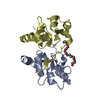
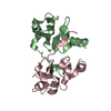
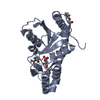
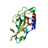
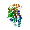
| Title | Crystal structure of a DUF4468 family protein (BACOVA_04320) from Bacteroides ovatus ATCC 8483 at 1.49 A resolution | ||||||
 Components Components | Uncharacterized protein | ||||||
 Keywords Keywords | UNKNOWN FUNCTION / PF14730 FAMILY PROTEIN / DUF4468 WITH TBP-LIKE FOLD /  STRUCTURAL GENOMICS / JOINT CENTER FOR STRUCTURAL GENOMICS / JCSG / PROTEIN STRUCTURE INITIATIVE / PSI-BIOLOGY STRUCTURAL GENOMICS / JOINT CENTER FOR STRUCTURAL GENOMICS / JCSG / PROTEIN STRUCTURE INITIATIVE / PSI-BIOLOGY | ||||||
| Function / homology | Alpha-D-Glucose-1,6-Bisphosphate; Chain A, domain 4 - #80 / Domain of unknown function DUF4468 with TBP-like fold / Domain of unknown function (DUF4468) with TBP-like fold / Alpha-D-Glucose-1,6-Bisphosphate; Chain A, domain 4 / 2-Layer Sandwich / Alpha Beta / NITRATE ION / DUF4468 domain-containing protein Function and homology information Function and homology information | ||||||
| Biological species |  Bacteroides ovatus (bacteria) Bacteroides ovatus (bacteria) | ||||||
| Method | X-RAY DIFFRACTION / SYNCHROTRON / MAD / Resolution: 1.49 Å | ||||||
 Authors Authors | Joint Center for Structural Genomics (JCSG) | ||||||
 Citation Citation | Journal: To be published Title: Crystal structure of a hypothetical protein (BACOVA_04320) from Bacteroides ovatus ATCC 8483 at 1.49 A resolution Authors: Joint Center for Structural Genomics (JCSG) | ||||||
| History |
|
- Structure visualization
| Structure viewer | Molecule: MolmilJmol/JSmol |
|---|
- Downloads & links
-Download
| PDBx/mmCIF format | 4e6f.cif.gz | 172.3 KB | Display | PDBx/mmCIF format |
|---|---|---|---|---|
| PDB format | pdb4e6f.ent.gz | 142.2 KB | Display | PDB format |
| PDBx/mmJSON format | 4e6f.json.gz | Tree view | PDBx/mmJSON format | |
| Others |  Other downloads Other downloads |
-Validation report
| Arichive directory | https://data.pdbj.org/pub/pdb/validation_reports/e6/4e6fftp://data.pdbj.org/pub/pdb/validation_reports/e6/4e6f | HTTPS FTP |
|---|
-Related structure data
| Similar structure data | |
|---|---|
| Other databases |


-Links
 PDBj
PDBj- Assembly
| Deposited unit | 
| ||||||||
|---|---|---|---|---|---|---|---|---|---|
| 1 | 
| ||||||||
| 2 | 
| ||||||||
| Unit cell |
|
-Components
| #1: Protein | Mass: 20934.107 Da / Num. of mol.: 2 / Fragment: UNP residues 25-384 Source method: isolated from a genetically manipulated source Source: (gene. exp.) Bacteroides ovatus (bacteria) / Gene: BACOVA_04320 / Plasmid: SpeedET / Production host: Escherichia coli (E. coli) / Strain (production host): PB1 / References: UniProt: A7M2I6#2: Chemical | ChemComp-NO3 / Nitrate  Mass: 62.005 Da / Num. of mol.: 7 / Source method: obtained synthetically / Formula: NO3 Mass: 62.005 Da / Num. of mol.: 7 / Source method: obtained synthetically / Formula: NO3#3: Chemical | Ethylene glycol  Mass: 62.068 Da / Num. of mol.: 3 / Source method: obtained synthetically / Formula: C2H6O2 Mass: 62.068 Da / Num. of mol.: 3 / Source method: obtained synthetically / Formula: C2H6O2#4: Water | ChemComp-HOH / | Water Mass: 18.015 Da / Num. of mol.: 721 / Source method: isolated from a natural source / Formula: H2O Mass: 18.015 Da / Num. of mol.: 721 / Source method: isolated from a natural source / Formula: H2OCompound details | THE UNIT CELL IS TOO SMALL TO CONTAIN THE INTACT PURIFIED PROTEIN CONSTRUCT RESIDUES 25-384). ...THE UNIT CELL IS TOO SMALL TO CONTAIN THE INTACT PURIFIED PROTEIN CONSTRUCT RESIDUES 25-384). THEREFORE, THE CRYSTAL MUST CONTAIN A PROTEOLYTI | Sequence details | THE CONSTRUCT WAS EXPRESSED WITH AN N-TERMINAL PURIFICATION TAG MGSDKIHHHHHHENLYFQG. THE TAG WAS ...THE CONSTRUCT WAS EXPRESSED WITH AN N-TERMINAL PURIFICATI | |
|---|
-Experimental details
-Experiment
| Experiment | Method: X-RAY DIFFRACTION / Number of used crystals: 1 |
|---|
- Sample preparation
| Crystal | Density Matthews: 2.65 Å3/Da / Density % sol: 53.66 % |
|---|---|
| Crystal grow | Temperature: 293 K / Method: vapor diffusion, sitting drop Details: 0.20M potassium nitrate, 20.00% polyethylene glycol 3350, NANODROP, VAPOR DIFFUSION, SITTING DROP, temperature 293K |
-Data collection
| Diffraction | Mean temperature: 100 K | |||||||||||||||||||||||||||||||||||||||||||||||||||||||||||||||||||||||||||||
|---|---|---|---|---|---|---|---|---|---|---|---|---|---|---|---|---|---|---|---|---|---|---|---|---|---|---|---|---|---|---|---|---|---|---|---|---|---|---|---|---|---|---|---|---|---|---|---|---|---|---|---|---|---|---|---|---|---|---|---|---|---|---|---|---|---|---|---|---|---|---|---|---|---|---|---|---|---|---|
| Diffraction source | Source: SYNCHROTRON / Site: SSRL  / Beamline: BL11-1 / Wavelength: 0.97972,0.91837,0.97917 / Beamline: BL11-1 / Wavelength: 0.97972,0.91837,0.97917 | |||||||||||||||||||||||||||||||||||||||||||||||||||||||||||||||||||||||||||||
| Detector | Type: DECTRIS PILATUS 6M / Detector: PIXEL / Date: Feb 8, 2012 Details: Flat mirror (vertical focusing); single crystal Si(111) bent monochromator (horizontal focusing) | |||||||||||||||||||||||||||||||||||||||||||||||||||||||||||||||||||||||||||||
| Radiation | Monochromator: single crystal Si(111) bent / Protocol: MAD / Monochromatic (M) / Laue (L): M / Scattering type: x-ray | |||||||||||||||||||||||||||||||||||||||||||||||||||||||||||||||||||||||||||||
| Radiation wavelength |
| |||||||||||||||||||||||||||||||||||||||||||||||||||||||||||||||||||||||||||||
| Reflection | Resolution: 1.49→47.449 Å / Num. obs: 69985 / % possible obs: 98.1 % / Observed criterion σ(I): -3 / Redundancy: 3.42 % / Biso Wilson estimate: 17.175 Å2 / Rmerge(I) obs: 0.054 / Net I/σ(I): 12.94 | |||||||||||||||||||||||||||||||||||||||||||||||||||||||||||||||||||||||||||||
| Reflection shell | Diffraction-ID: 1
|
-Phasing
| Phasing | Method: MAD |
|---|
- Processing
| Software |
| ||||||||||||||||||||||||||||||||||||||||||||||||||||||||||||||||||||||||||||||||||||||||||||||||||||||||||||
|---|---|---|---|---|---|---|---|---|---|---|---|---|---|---|---|---|---|---|---|---|---|---|---|---|---|---|---|---|---|---|---|---|---|---|---|---|---|---|---|---|---|---|---|---|---|---|---|---|---|---|---|---|---|---|---|---|---|---|---|---|---|---|---|---|---|---|---|---|---|---|---|---|---|---|---|---|---|---|---|---|---|---|---|---|---|---|---|---|---|---|---|---|---|---|---|---|---|---|---|---|---|---|---|---|---|---|---|---|---|
| Refinement | Method to determine structure: MAD / Resolution: 1.49→47.449 Å / Cor.coef. Fo:Fc: 0.9677 / Cor.coef. Fo:Fc free: 0.9604 / Occupancy max: 1 / Occupancy min: 0.25 / Cross valid method: THROUGHOUT / σ(F): 0 Details: 1. A MET-INHIBITION PROTOCOL WAS USED FOR SELENOMETHIONINE INCORPORATION DURING PROTEIN EXPRESSION. THE OCCUPANCY OF THE SE ATOMS IN THE MSE RESIDUES WAS REDUCED TO 0.75 FOR THE REDUCED ...Details: 1. A MET-INHIBITION PROTOCOL WAS USED FOR SELENOMETHIONINE INCORPORATION DURING PROTEIN EXPRESSION. THE OCCUPANCY OF THE SE ATOMS IN THE MSE RESIDUES WAS REDUCED TO 0.75 FOR THE REDUCED SCATTERING POWER DUE TO PARTIAL S-MET INCORPORATION. 2. ATOM RECORD CONTAINS SUM OF TLS AND RESIDUAL B FACTORS. ANISOU RECORD CONTAINS SUM OF TLS AND RESIDUAL U FACTORS. 3. THE MAD PHASES WERE USED AS RESTRAINTS DURING REFINEMENT. 4. NITRATE AND ETHYLENE GLYCOL MODELED WERE PRESENT IN CRYSTALLIZATION CONDITION OR CRYOPROTECTANT. 5. THE C-TERMINAL PORTION OF THE PROTEIN WAS PRESENT AFTER PURIFICATION, BUT IS NOT OBSERVED IN THE CRYSTAL STRUCTURE. THE SIZE OF THE UNIT CELL IS TOO SMALL TO CONTAIN THE FULL CONSTRUCT. THEREFORE THE CRYSTAL MUST CONTAIN A PROTEOLYTIC FRAGMENT. HOWEVER, THE SITE OF PROTEOLYSIS IS UNKNOWN. RESIDUES 30-200 WERE MODELED IN CHAIN A AND 32-204 IN CHAIN B.
| ||||||||||||||||||||||||||||||||||||||||||||||||||||||||||||||||||||||||||||||||||||||||||||||||||||||||||||
| Displacement parameters | Biso max: 106.26 Å2 / Biso mean: 24.53 Å2 / Biso min: 7.17 Å2
| ||||||||||||||||||||||||||||||||||||||||||||||||||||||||||||||||||||||||||||||||||||||||||||||||||||||||||||
| Refine analyze | Luzzati coordinate error obs: 0.18 Å | ||||||||||||||||||||||||||||||||||||||||||||||||||||||||||||||||||||||||||||||||||||||||||||||||||||||||||||
| Refinement step | Cycle: LAST / Resolution: 1.49→47.449 Å
| ||||||||||||||||||||||||||||||||||||||||||||||||||||||||||||||||||||||||||||||||||||||||||||||||||||||||||||
| Refine LS restraints |
| ||||||||||||||||||||||||||||||||||||||||||||||||||||||||||||||||||||||||||||||||||||||||||||||||||||||||||||
| LS refinement shell | Resolution: 1.49→1.53 Å / Total num. of bins used: 20
| ||||||||||||||||||||||||||||||||||||||||||||||||||||||||||||||||||||||||||||||||||||||||||||||||||||||||||||
| Refinement TLS params. | Method: refined / Refine-ID: X-RAY DIFFRACTION
| ||||||||||||||||||||||||||||||||||||||||||||||||||||||||||||||||||||||||||||||||||||||||||||||||||||||||||||
| Refinement TLS group |
|
