[English] 日本語
Yorodumi
- PDB-4e36: Crystal structure of the human Endoplasmic Reticulum Aminopeptida... -

+
Open data


ID or keywords:

Loading...

-
Basic information

Entry
Database: PDB / ID: 4.0E+36
TitleCrystal structure of the human Endoplasmic Reticulum Aminopeptidase 2 variant N392K
ComponentsEndoplasmic reticulum aminopeptidase 2
Keywords
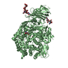
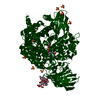
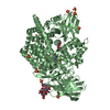
HYDROLASE / Thermolysin-like catalytic domain / Aminopeptidase / Zinc binding / Glycosylation / Endoplasmic Reticulum
Function / homology
Function and homology information


Hydrolases; Acting on peptide bonds (peptidases); Aminopeptidases / antigen processing and presentation of peptide antigen via MHC class I / peptide catabolic process / metalloaminopeptidase activity / aminopeptidase activity / Antigen Presentation: Folding, assembly and peptide loading of class I MHC / peptide binding / antigen processing and presentation of endogenous peptide antigen via MHC class I / regulation of blood pressure / metallopeptidase activity ...Hydrolases; Acting on peptide bonds (peptidases); Aminopeptidases / antigen processing and presentation of peptide antigen via MHC class I / peptide catabolic process / metalloaminopeptidase activity / aminopeptidase activity / Antigen Presentation: Folding, assembly and peptide loading of class I MHC / peptide binding / antigen processing and presentation of endogenous peptide antigen via MHC class I / regulation of blood pressure / metallopeptidase activity / endopeptidase activity / adaptive immune response / endoplasmic reticulum lumen / endoplasmic reticulum membrane / proteolysis / extracellular space / zinc ion binding / membrane / cytoplasm
Similarity search - Function
Zincin-like fold - #20 / Immunoglobulin-like - #1910 / Aminopeptidase N-type / ERAP1-like C-terminal domain / ERAP1-like C-terminal domain / Zincin-like fold / tricorn interacting facor f3 domain / Aminopeptidase N-like , N-terminal domain / Peptidase M1, alanine aminopeptidase/leukotriene A4 hydrolase / Peptidase M1, membrane alanine aminopeptidase ...Zincin-like fold - #20 / Immunoglobulin-like - #1910 / Aminopeptidase N-type / ERAP1-like C-terminal domain / ERAP1-like C-terminal domain / Zincin-like fold / tricorn interacting facor f3 domain / Aminopeptidase N-like , N-terminal domain / Peptidase M1, alanine aminopeptidase/leukotriene A4 hydrolase / Peptidase M1, membrane alanine aminopeptidase / Peptidase family M1 domain / Peptidase M1 N-terminal domain / Aminopeptidase N-like , N-terminal domain superfamliy / Neutral Protease Domain 2 / Neutral Protease; domain 2 / Peptidase M4/M1, CTD superfamily / Neutral zinc metallopeptidases, zinc-binding region signature. / Alpha Horseshoe / Immunoglobulin-like / Sandwich / Orthogonal Bundle / Mainly Beta / Mainly Alpha
Similarity search - Domain/homology
LYSINE / Endoplasmic reticulum aminopeptidase 2
Similarity search - Component
Biological speciesHomo sapiens (human)
MethodX-RAY DIFFRACTION / SYNCHROTRON / isomorphous replacement / Resolution: 3.22 Å
AuthorsBirtley, J.R. / Saridakis, E. / Pegias, P. / Stratikos, E. / Mavridis, I.M.
CitationJournal: J.Immunol. / Year: 2012
Title: A common single nucleotide polymorphism in endoplasmic reticulum aminopeptidase 2 induces a specificity switch that leads to altered antigen processing.
Authors: Evnouchidou, I. / Birtley, J. / Seregin, S. / Papakyriakou, A. / Zervoudi, E. / Samiotaki, M. / Panayotou, G. / Giastas, P. / Petrakis, O. / Georgiadis, D. / Amalfitano, A. / Saridakis, E. / ...Authors: Evnouchidou, I. / Birtley, J. / Seregin, S. / Papakyriakou, A. / Zervoudi, E. / Samiotaki, M. / Panayotou, G. / Giastas, P. / Petrakis, O. / Georgiadis, D. / Amalfitano, A. / Saridakis, E. / Mavridis, I.M. / Stratikos, E.
History
DepositionMar 9, 2012Deposition site: RCSB / Processing site: RCSB
Revision 1.0Aug 29, 2012Provider: repository / Type: Initial release
Revision 1.1Oct 17, 2012Group: Database references
Revision 1.2Nov 15, 2017Group: Advisory / Refinement description / Category: pdbx_unobs_or_zero_occ_atoms / software
Revision 2.0Jul 29, 2020Group: Advisory / Atomic model ...Advisory / Atomic model / Data collection / Database references / Derived calculations / Structure summary
Category: atom_site / chem_comp ...atom_site / chem_comp / entity / pdbx_branch_scheme / pdbx_chem_comp_identifier / pdbx_entity_branch / pdbx_entity_branch_descriptor / pdbx_entity_branch_link / pdbx_entity_branch_list / pdbx_entity_nonpoly / pdbx_nonpoly_scheme / pdbx_struct_assembly_gen / pdbx_struct_conn_angle / pdbx_validate_close_contact / struct_asym / struct_conn / struct_ref_seq_dif / struct_site / struct_site_gen
Item: _atom_site.B_iso_or_equiv / _atom_site.Cartn_x ..._atom_site.B_iso_or_equiv / _atom_site.Cartn_x / _atom_site.Cartn_y / _atom_site.Cartn_z / _atom_site.auth_asym_id / _atom_site.auth_atom_id / _atom_site.auth_comp_id / _atom_site.auth_seq_id / _atom_site.label_asym_id / _atom_site.label_atom_id / _atom_site.label_comp_id / _atom_site.label_entity_id / _atom_site.occupancy / _atom_site.type_symbol / _chem_comp.name / _chem_comp.type / _pdbx_struct_assembly_gen.asym_id_list / _pdbx_struct_conn_angle.ptnr1_auth_asym_id / _pdbx_struct_conn_angle.ptnr1_auth_comp_id / _pdbx_struct_conn_angle.ptnr1_auth_seq_id / _pdbx_struct_conn_angle.ptnr1_label_asym_id / _pdbx_struct_conn_angle.ptnr1_label_atom_id / _pdbx_struct_conn_angle.ptnr1_label_comp_id / _pdbx_struct_conn_angle.ptnr1_label_seq_id / _pdbx_struct_conn_angle.ptnr2_auth_asym_id / _pdbx_struct_conn_angle.ptnr2_auth_seq_id / _pdbx_struct_conn_angle.ptnr2_label_asym_id / _pdbx_struct_conn_angle.ptnr3_auth_asym_id / _pdbx_struct_conn_angle.ptnr3_auth_comp_id / _pdbx_struct_conn_angle.ptnr3_auth_seq_id / _pdbx_struct_conn_angle.ptnr3_label_asym_id / _pdbx_struct_conn_angle.ptnr3_label_atom_id / _pdbx_struct_conn_angle.ptnr3_label_comp_id / _pdbx_struct_conn_angle.ptnr3_label_seq_id / _pdbx_struct_conn_angle.value / _pdbx_validate_close_contact.auth_asym_id_1 / _pdbx_validate_close_contact.auth_asym_id_2 / _pdbx_validate_close_contact.auth_seq_id_1 / _pdbx_validate_close_contact.auth_seq_id_2 / _struct_conn.pdbx_dist_value / _struct_conn.pdbx_leaving_atom_flag / _struct_conn.pdbx_role / _struct_conn.ptnr1_auth_asym_id / _struct_conn.ptnr1_auth_comp_id / _struct_conn.ptnr1_auth_seq_id / _struct_conn.ptnr1_label_asym_id / _struct_conn.ptnr1_label_atom_id / _struct_conn.ptnr1_label_comp_id / _struct_conn.ptnr1_label_seq_id / _struct_conn.ptnr2_auth_asym_id / _struct_conn.ptnr2_auth_comp_id / _struct_conn.ptnr2_auth_seq_id / _struct_conn.ptnr2_label_asym_id / _struct_conn.ptnr2_label_atom_id / _struct_conn.ptnr2_label_comp_id / _struct_ref_seq_dif.details
Description: Carbohydrate remediation / Provider: repository / Type: Remediation
Revision 2.1Sep 13, 2023Group: Advisory / Data collection ...Advisory / Data collection / Database references / Refinement description / Structure summary
Category: chem_comp / chem_comp_atom ...chem_comp / chem_comp_atom / chem_comp_bond / database_2 / pdbx_initial_refinement_model / pdbx_unobs_or_zero_occ_atoms
Item: _chem_comp.pdbx_synonyms / _database_2.pdbx_DOI / _database_2.pdbx_database_accession

-
Structure visualization

Structure viewerMolecule:
MolmilJmol/JSmol

Downloads & links

-
Assembly

Deposited unit
A: Endoplasmic reticulum aminopeptidase 2
B: Endoplasmic reticulum aminopeptidase 2
hetero molecules


Theoretical massNumber of molelcules
Total (without water)226,78316
Polymers223,0612
Non-polymers3,72214
Water1,910106
1
A: Endoplasmic reticulum aminopeptidase 2
hetero molecules


Theoretical massNumber of molelcules
Total (without water)113,4519
Polymers111,5301
Non-polymers1,9208
Water181
TypeNameSymmetry operationNumber
identity operation1_555x,y,z1
MethodPISA
2
B: Endoplasmic reticulum aminopeptidase 2
hetero molecules


Theoretical massNumber of molelcules
Total (without water)113,3337
Polymers111,5301
Non-polymers1,8026
Water181
TypeNameSymmetry operationNumber
identity operation1_555x,y,z1
MethodPISA
Unit cell
Length a, b, c (Å)74.340, 134.450, 127.370
Angle α, β, γ (deg.)90.00, 90.85, 90.00
Int Tables number4
Space group name H-MP1211
Noncrystallographic symmetry (NCS)NCS domain:
IDEns-ID
11
21

NCS domain segments:
Dom-IDComponent-IDEns-IDSelection details
111chain A and (resseq 61:125 or resseq 134:500 or resseq...
211chain B and (resseq 61:125 or resseq 134:500 or resseq...

-
Components

-
Protein , 1 types, 2 molecules AB

#1: Protein Endoplasmic reticulum aminopeptidase 2 / Leukocyte-derived arginine aminopeptidase / L-RAP


Mass: 111530.453 Da / Num. of mol.: 2
Source method: isolated from a genetically manipulated source
Source: (gene. exp.) Homo sapiens (human) / Gene: ERAP2, Irap, LRAP / Plasmid: pFASTBac-1 / Production host: Trichoplusia ni (cabbage looper)
References: UniProt: Q6P179, Hydrolases; Acting on peptide bonds (peptidases); Aminopeptidases

-
Sugars , 3 types, 8 molecules

#2: Polysaccharide 2-acetamido-2-deoxy-beta-D-glucopyranose-(1-4)-2-acetamido-2-deoxy-beta-D-glucopyranose


Type: oligosaccharide / Mass: 424.401 Da / Num. of mol.: 3
Source method: isolated from a genetically manipulated source
DescriptorTypeProgram
DGlcpNAcb1-4DGlcpNAcb1-Glycam Condensed SequenceGMML 1.0
WURCS=2.0/1,2,1/[a2122h-1b_1-5_2*NCC/3=O]/1-1/a4-b1WURCSPDB2Glycan 1.1.0
[]{[(4+1)][b-D-GlcpNAc]{[(4+1)][b-D-GlcpNAc]{}}}LINUCSPDB-CARE
#3: Polysaccharide alpha-D-mannopyranose-(1-4)-alpha-D-mannopyranose-(1-4)-2-acetamido-2-deoxy-beta-D-glucopyranose-(1- ...alpha-D-mannopyranose-(1-4)-alpha-D-mannopyranose-(1-4)-2-acetamido-2-deoxy-beta-D-glucopyranose-(1-4)-2-acetamido-2-deoxy-beta-D-glucopyranose


Type: oligosaccharide / Mass: 748.682 Da / Num. of mol.: 1
Source method: isolated from a genetically manipulated source
DescriptorTypeProgram
DManpa1-4DManpa1-4DGlcpNAcb1-4DGlcpNAcb1-Glycam Condensed SequenceGMML 1.0
WURCS=2.0/2,4,3/[a2122h-1b_1-5_2*NCC/3=O][a1122h-1a_1-5]/1-1-2-2/a4-b1_b4-c1_c4-d1WURCSPDB2Glycan 1.1.0
[]{[(4+1)][b-D-GlcpNAc]{[(4+1)][b-D-GlcpNAc]{[(4+1)][a-D-Manp]{[(4+1)][a-D-Manp]{}}}}}LINUCSPDB-CARE
#5: Sugar
ChemComp-NAG / 2-acetamido-2-deoxy-beta-D-glucopyranose / N-acetyl-beta-D-glucosamine / 2-acetamido-2-deoxy-beta-D-glucose / 2-acetamido-2-deoxy-D-glucose / 2-acetamido-2-deoxy-glucose / N-ACETYL-D-GLUCOSAMINE / N-Acetylglucosamine


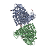
Type: D-saccharide, beta linking / Mass: 221.208 Da / Num. of mol.: 4
Source method: isolated from a genetically manipulated source
Formula: C8H15NO6
IdentifierTypeProgram
DGlcpNAcbCONDENSED IUPAC CARBOHYDRATE SYMBOLGMML 1.0
N-acetyl-b-D-glucopyranosamineCOMMON NAMEGMML 1.0
b-D-GlcpNAcIUPAC CARBOHYDRATE SYMBOLPDB-CARE 1.0
GlcNAcSNFG CARBOHYDRATE SYMBOLGMML 1.0

-
Non-polymers , 4 types, 112 molecules

#4: Chemical ChemComp-LYS / LYSINE / Lysine


Type: L-peptide linking / Mass: 147.195 Da / Num. of mol.: 2 / Source method: obtained synthetically / Formula: C6H15N2O2
#6: Chemical ChemComp-MES / 2-(N-MORPHOLINO)-ETHANESULFONIC ACID / MES (buffer)


Mass: 195.237 Da / Num. of mol.: 2 / Source method: obtained synthetically / Formula: C6H13NO4S / Comment: pH buffer*YM
#7: Chemical ChemComp-ZN / ZINC ION


Mass: 65.409 Da / Num. of mol.: 2 / Source method: obtained synthetically / Formula: Zn
#8: Water ChemComp-HOH / water / Water


Mass: 18.015 Da / Num. of mol.: 106 / Source method: isolated from a natural source / Formula: H2O

-
Details

Sequence detailsTHE DISCREPANCY BETWEEN THE UNIPPROT ENTRY AND THE PROVIDED SEQUENCE AT RESIDUE 2 CORRESPONDS TO A ...THE DISCREPANCY BETWEEN THE UNIPPROT ENTRY AND THE PROVIDED SEQUENCE AT RESIDUE 2 CORRESPONDS TO A COMMON CLONING ARTIFACT.

-
Experimental details

-
Experiment

ExperimentMethod: X-RAY DIFFRACTION / Number of used crystals: 1

-
Sample preparation

CrystalDensity Matthews: 2.85 Å3/Da / Density % sol: 56.86 %
Crystal growTemperature: 277 K / Method: vapor diffusion, hanging drop / pH: 6.5
Details: 10% PEG 8000, 20% ethylene glycol, 20 mM glycine, 20 mM DL-lysine, 20 mM DL-serine, 20 mM DL-alanine, 20 mM sodium L-glutamate, 31 mM imidazole, 69 mM MES. Protein mixed 1:4 with LPL ...Details: 10% PEG 8000, 20% ethylene glycol, 20 mM glycine, 20 mM DL-lysine, 20 mM DL-serine, 20 mM DL-alanine, 20 mM sodium L-glutamate, 31 mM imidazole, 69 mM MES. Protein mixed 1:4 with LPL peptide, pH 6.5, VAPOR DIFFUSION, HANGING DROP, temperature 277K

-
Data collection

DiffractionMean temperature: 100 K
Diffraction sourceSource: SYNCHROTRON / Site: SLS / Beamline: X06DA / Wavelength: 1 Å
DetectorType: DECTRIS PILATUS 2M / Detector: PIXEL / Date: Dec 4, 2011
RadiationMonochromator: BARTELS / Protocol: SINGLE WAVELENGTH / Monochromatic (M) / Laue (L): M / Scattering type: x-ray
Radiation wavelengthWavelength: 1 Å / Relative weight: 1
ReflectionResolution: 3.22→59.45 Å / Num. obs: 71589 / % possible obs: 99.1 % / Observed criterion σ(I): -5
Reflection shellResolution: 3.22→3.39 Å / % possible all: 99.39

-
Processing

Software
NameVersionClassification
MOLREPphasing
PHENIX(phenix.refine)refinement
MOSFLMdata reduction
SCALAdata scaling
RefinementMethod to determine structure: isomorphous replacement
Starting model: PDB ENTRY 3SE6
Resolution: 3.22→10.996 Å / σ(F): 1.12 / Phase error: 28.72 / Stereochemistry target values: TWIN_LSQ_F
RfactorNum. reflection% reflection
Rfree0.2608 3912 5.62 %
Rwork0.2087 --
obs0.2113 69634 89.42 %
all-77873 -
Solvent computationShrinkage radii: 0.9 Å / VDW probe radii: 1.11 Å / Solvent model: FLAT BULK SOLVENT MODEL / Bsol: 55.818 Å2 / ksol: 0.312 e/Å3
Displacement parameters
Baniso -1Baniso -2Baniso -3
1-5.1882 Å2-0 Å215.2206 Å2
2---15.5805 Å2-0 Å2
3---6.3347 Å2
Refinement stepCycle: LAST / Resolution: 3.22→10.996 Å
ProteinNucleic acidLigandSolventTotal
Num. atoms13985 0 236 106 14327
Refine LS restraints
Refine-IDTypeDev idealNumber
X-RAY DIFFRACTIONf_bond_d0.00714600
X-RAY DIFFRACTIONf_angle_d1.1219779
X-RAY DIFFRACTIONf_dihedral_angle_d19.5055307
X-RAY DIFFRACTIONf_chiral_restr0.0652227
X-RAY DIFFRACTIONf_plane_restr0.0042450
Refine LS restraints NCS
Ens-IDDom-IDAuth asym-IDNumberRefine-IDTypeRms dev position (Å)
11A6804X-RAY DIFFRACTIONPOSITIONAL
12B6804X-RAY DIFFRACTIONPOSITIONAL0.037
LS refinement shell
Resolution (Å)Rfactor RfreeNum. reflection RfreeRfactor RworkNum. reflection RworkRefine-ID% reflection obs (%)
3.22-3.27410.37211470.27873443X-RAY DIFFRACTION98
3.2741-3.3320.3351540.27483406X-RAY DIFFRACTION98
3.332-3.39430.34431720.28023442X-RAY DIFFRACTION98
3.3943-3.46140.30131760.25453366X-RAY DIFFRACTION98
3.4614-3.53430.30762060.25913382X-RAY DIFFRACTION98
3.5343-3.61360.32611860.24023283X-RAY DIFFRACTION98
3.6136-3.70060.34261870.23693331X-RAY DIFFRACTION98
3.7006-3.79670.26791690.21873237X-RAY DIFFRACTION98
3.7967-3.90360.25231760.22273224X-RAY DIFFRACTION98
3.9036-4.02360.25091750.22292962X-RAY DIFFRACTION98
4.0236-4.15990.29291700.21573035X-RAY DIFFRACTION98
4.1599-4.31670.28031440.19863227X-RAY DIFFRACTION98
4.3167-4.50030.24251430.18773336X-RAY DIFFRACTION98
4.5003-4.71970.25461880.19243481X-RAY DIFFRACTION98
4.7197-4.98920.21731830.18253430X-RAY DIFFRACTION98
4.9892-5.33330.25521990.19683343X-RAY DIFFRACTION98
5.3333-5.79750.27661550.20193412X-RAY DIFFRACTION98
5.7975-6.48190.22791660.21563376X-RAY DIFFRACTION98
6.4819-7.67490.251650.18883151X-RAY DIFFRACTION98
7.6749-10.99650.17382090.14663297X-RAY DIFFRACTION98

+
About Yorodumi

-
News

-
Feb 9, 2022. New format data for meta-information of EMDB entries

New format data for meta-information of EMDB entries

  • Version 3 of the EMDB header file is now the official format.
  • The previous official version 1.9 will be removed from the archive.

Related info.:EMDB header

External links:wwPDB to switch to version 3 of the EMDB data model

-
Aug 12, 2020. Covid-19 info

Covid-19 info

URL: https://pdbj.org/emnavi/covid19.php

New page: Covid-19 featured information page in EM Navigator.

Related info.:Covid-19 info / Mar 5, 2020. Novel coronavirus structure data

+
Mar 5, 2020. Novel coronavirus structure data

Novel coronavirus structure data

Related info.:Yorodumi Speices / Aug 12, 2020. Covid-19 info

External links:COVID-19 featured content - PDBj / Molecule of the Month (242):Coronavirus Proteases

+
Jan 31, 2019. EMDB accession codes are about to change! (news from PDBe EMDB page)

EMDB accession codes are about to change! (news from PDBe EMDB page)

  • The allocation of 4 digits for EMDB accession codes will soon come to an end. Whilst these codes will remain in use, new EMDB accession codes will include an additional digit and will expand incrementally as the available range of codes is exhausted. The current 4-digit format prefixed with “EMD-” (i.e. EMD-XXXX) will advance to a 5-digit format (i.e. EMD-XXXXX), and so on. It is currently estimated that the 4-digit codes will be depleted around Spring 2019, at which point the 5-digit format will come into force.
  • The EM Navigator/Yorodumi systems omit the EMD- prefix.

Related info.:Q: What is EMD? / ID/Accession-code notation in Yorodumi/EM Navigator

External links:EMDB Accession Codes are Changing Soon! / Contact to PDBj

+
Jul 12, 2017. Major update of PDB

Major update of PDB

  • wwPDB released updated PDB data conforming to the new PDBx/mmCIF dictionary.
  • This is a major update changing the version number from 4 to 5, and with Remediation, in which all the entries are updated.
  • In this update, many items about electron microscopy experimental information are reorganized (e.g. em_software).
  • Now, EM Navigator and Yorodumi are based on the updated data.

External links:wwPDB Remediation / Enriched Model Files Conforming to OneDep Data Standards Now Available in the PDB FTP Archive

-
Yorodumi

Thousand views of thousand structures

  • Yorodumi is a browser for structure data from EMDB, PDB, SASBDB, etc.
  • This page is also the successor to EM Navigator detail page, and also detail information page/front-end page for Omokage search.
  • The word "yorodu" (or yorozu) is an old Japanese word meaning "ten thousand". "mi" (miru) is to see.

Related info.:EMDB / PDB / SASBDB / Comparison of 3 databanks / Yorodumi Search / Aug 31, 2016. New EM Navigator & Yorodumi / Yorodumi Papers / Jmol/JSmol / Function and homology information / Changes in new EM Navigator and Yorodumi

Read more