 Movie
Movie Controller

[English] 日本語
 Yorodumi
- PDB-4e0f: Crystallographic structure of trimeric Riboflavin Synthase from B... -
+ Open data

- Basic information
| Entry | Database: PDB / ID: 4e0f | ||||||
|---|---|---|---|---|---|---|---|
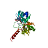
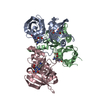
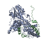
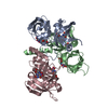
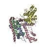
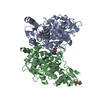
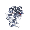
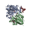
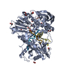
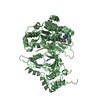
| Title | Crystallographic structure of trimeric Riboflavin Synthase from Brucella abortus in complex with riboflavin | ||||||
 Components Components | Riboflavin synthase subunit alpha | ||||||
 Keywords Keywords | TRANSFERASE / beta barrel / alpha + beta protein / Riboflavin biosynthesis | ||||||
| Function / homology | Elongation Factor Tu (Ef-tu); domain 3 - #20 / Elongation Factor Tu (Ef-tu); domain 3 / Beta Barrel / Mainly Beta / RIBOFLAVIN / :  Function and homology information Function and homology information | ||||||
| Biological species |  Brucella abortus (bacteria) Brucella abortus (bacteria) | ||||||
| Method | X-RAY DIFFRACTION / SYNCHROTRON / MOLECULAR REPLACEMENT / Resolution: 2.85 Å | ||||||
 Authors Authors | Serer, M.I. / Bonomi, H.R. / Guimaraes, B.G. / Rossi, R.C. / Goldbaum, F.A. / Klinke, S. | ||||||
 Citation Citation | Journal: Acta Crystallogr.,Sect.D / Year: 2014 Title: Crystallographic and kinetic study of riboflavin synthase from Brucella abortus, a chemotherapeutic target with an enhanced intrinsic flexibility. Authors: Serer, M.I. / Bonomi, H.R. / Guimaraes, B.G. / Rossi, R.C. / Goldbaum, F.A. / Klinke, S. | ||||||
| History |
|
- Structure visualization
| Structure viewer | Molecule: MolmilJmol/JSmol |
|---|
- Downloads & links
-Download
| PDBx/mmCIF format | 4e0f.cif.gz | 125.2 KB | Display | PDBx/mmCIF format |
|---|---|---|---|---|
| PDB format | pdb4e0f.ent.gz | 97.2 KB | Display | PDB format |
| PDBx/mmJSON format | 4e0f.json.gz | Tree view | PDBx/mmJSON format | |
| Others |  Other downloads Other downloads |
-Validation report
| Arichive directory | https://data.pdbj.org/pub/pdb/validation_reports/e0/4e0fftp://data.pdbj.org/pub/pdb/validation_reports/e0/4e0f | HTTPS FTP |
|---|
-Related structure data
| Related structure data | 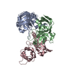 4fxuC  4g6iC  4gqnC 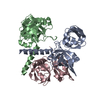 1i8dS S: Starting model for refinement C: citing same article ( |
|---|---|
| Similar structure data |

-Links
 PDBj
PDBj- Assembly
| Deposited unit | 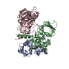
| ||||||||
|---|---|---|---|---|---|---|---|---|---|
| 1 |
| ||||||||
| Unit cell |
|
-Components
| #1: Protein | Mass: 23320.361 Da / Num. of mol.: 3 Source method: isolated from a genetically manipulated source Source: (gene. exp.) Brucella abortus (bacteria) / Gene: ribE / Plasmid: pET22b / Production host: Escherichia coli (E. coli) / Strain (production host): BL21 (DE3) / References: UniProt: G8SX20, riboflavin synthase#2: Chemical | ChemComp-RBF / | Riboflavin  Mass: 376.364 Da / Num. of mol.: 1 / Source method: obtained synthetically / Formula: C17H20N4O6 Mass: 376.364 Da / Num. of mol.: 1 / Source method: obtained synthetically / Formula: C17H20N4O6#3: Water | ChemComp-HOH / | Water Mass: 18.015 Da / Num. of mol.: 66 / Source method: isolated from a natural source / Formula: H2O Mass: 18.015 Da / Num. of mol.: 66 / Source method: isolated from a natural source / Formula: H2O |
|---|
-Experimental details
-Experiment
| Experiment | Method: X-RAY DIFFRACTION / Number of used crystals: 1 |
|---|
- Sample preparation
| Crystal | Density Matthews: 2.27 Å3/Da / Density % sol: 45.76 % |
|---|---|
| Crystal grow | Temperature: 292 K / Method: vapor diffusion, hanging drop / pH: 7.4 Details: 12% PEG 8000, 10% Glycerol, 0.5M Potassium Chloride, pH 7.4, VAPOR DIFFUSION, HANGING DROP, temperature 292K |
-Data collection
| Diffraction | Mean temperature: 100 K |
|---|---|
| Diffraction source | Source: SYNCHROTRON / Site: NSLS  / Beamline: X6A / Wavelength: 1 Å / Beamline: X6A / Wavelength: 1 Å |
| Detector | Type: ADSC QUANTUM 270 / Detector: CCD / Date: Oct 8, 2011 / Details: Toroidal focusing mirror |
| Radiation | Monochromator: Si(111) channel cut monochromator / Protocol: SINGLE WAVELENGTH / Monochromatic (M) / Laue (L): M / Scattering type: x-ray |
| Radiation wavelength | Wavelength: 1 Å / Relative weight: 1 |
| Reflection | Resolution: 2.85→67.25 Å / Num. all: 15167 / Num. obs: 15167 / % possible obs: 98.7 % / Observed criterion σ(F): 0 / Observed criterion σ(I): 0 / Redundancy: 8.6 % / Biso Wilson estimate: 47.8 Å2 / Rmerge(I) obs: 0.123 / Rsym value: 0.123 / Net I/σ(I): 5.9 |
| Reflection shell | Resolution: 2.85→3 Å / Redundancy: 8.7 % / Rmerge(I) obs: 0.385 / Mean I/σ(I) obs: 1.9 / Num. unique all: 2177 / Rsym value: 0.385 / % possible all: 98.6 |
- Processing
| Software |
| |||||||||||||||||||||||||||||||||||||||||||||||||||||||||||||||||
|---|---|---|---|---|---|---|---|---|---|---|---|---|---|---|---|---|---|---|---|---|---|---|---|---|---|---|---|---|---|---|---|---|---|---|---|---|---|---|---|---|---|---|---|---|---|---|---|---|---|---|---|---|---|---|---|---|---|---|---|---|---|---|---|---|---|---|
| Refinement | Method to determine structure: MOLECULAR REPLACEMENT Starting model: Monomer from PDB entry 1I8D Resolution: 2.85→67.25 Å / Cor.coef. Fo:Fc: 0.897 / Cor.coef. Fo:Fc free: 0.829 / SU B: 16.354 / SU ML: 0.32 / Cross valid method: THROUGHOUT / ESU R Free: 0.445 / Stereochemistry target values: MAXIMUM LIKELIHOOD
| |||||||||||||||||||||||||||||||||||||||||||||||||||||||||||||||||
| Solvent computation | Ion probe radii: 0.8 Å / Shrinkage radii: 0.8 Å / VDW probe radii: 1.4 Å / Solvent model: MASK | |||||||||||||||||||||||||||||||||||||||||||||||||||||||||||||||||
| Displacement parameters | Biso mean: 22.521 Å2
| |||||||||||||||||||||||||||||||||||||||||||||||||||||||||||||||||
| Refinement step | Cycle: LAST / Resolution: 2.85→67.25 Å
| |||||||||||||||||||||||||||||||||||||||||||||||||||||||||||||||||
| Refine LS restraints |
| |||||||||||||||||||||||||||||||||||||||||||||||||||||||||||||||||
| LS refinement shell | Resolution: 2.85→2.924 Å / Total num. of bins used: 20
|
