 Movie
Movie Controller

[English] 日本語
 Yorodumi
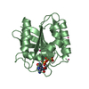
- PDB-4dzr: The crystal structure of protein-(glutamine-N5) methyltransferase... -
+ Open data

- Basic information
| Entry | Database: PDB / ID: 4dzr | ||||||
|---|---|---|---|---|---|---|---|
| Title | The crystal structure of protein-(glutamine-N5) methyltransferase (release factor-specific) from Alicyclobacillus acidocaldarius subsp. acidocaldarius DSM 446 | ||||||
 Components Components | Protein-(Glutamine-N5) methyltransferase, release factor-specific | ||||||
 Keywords Keywords |  TRANSFERASE / structural genomics / PSI-Biology / protein structure initiative / midwest center for structural genomics / MCSG TRANSFERASE / structural genomics / PSI-Biology / protein structure initiative / midwest center for structural genomics / MCSG | ||||||
| Function / homology |  Function and homology information Function and homology informationpeptide chain release factor N5-glutamine methyltransferase / protein-(glutamine-N5) methyltransferase activity / protein-glutamine N-methyltransferase activity / peptidyl-glutamine methylation / nucleic acid binding / metal ion bindingSimilarity search - Function | ||||||
| Biological species |  Alicyclobacillus acidocaldarius subsp. acidocaldarius DSM 446 (bacteria) Alicyclobacillus acidocaldarius subsp. acidocaldarius DSM 446 (bacteria) | ||||||
| Method | X-RAY DIFFRACTION / SYNCHROTRON / MAD / Resolution: 2.551 Å | ||||||
 Authors Authors | Tan, K. / Chhor, G. / Bearden, J. / Joachimiak, A. / Midwest Center for Structural Genomics (MCSG) | ||||||
 Citation Citation | Journal: To be Published Title: The crystal structure of protein-(glutamine-N5) methyltransferase (release factor-specific) from Alicyclobacillus acidocaldarius subsp. acidocaldarius DSM 446 Authors: Tan, K. / Chhor, G. / Bearden, J. / Joachimiak, A. | ||||||
| History |
|
- Structure visualization
| Structure viewer | Molecule: MolmilJmol/JSmol |
|---|
- Downloads & links
-Download
| PDBx/mmCIF format | 4dzr.cif.gz | 76.9 KB | Display | PDBx/mmCIF format |
|---|---|---|---|---|
| PDB format | pdb4dzr.ent.gz | 61.2 KB | Display | PDB format |
| PDBx/mmJSON format | 4dzr.json.gz | Tree view | PDBx/mmJSON format | |
| Others |  Other downloads Other downloads |
-Validation report
| Arichive directory | https://data.pdbj.org/pub/pdb/validation_reports/dz/4dzrftp://data.pdbj.org/pub/pdb/validation_reports/dz/4dzr | HTTPS FTP |
|---|
-Related structure data
| Similar structure data | |
|---|---|
| Other databases |

-Links
 PDBj
PDBj- Assembly
| Deposited unit | 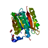
| |||||||||
|---|---|---|---|---|---|---|---|---|---|---|
| 1 |
| |||||||||
| 2 | 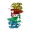
| |||||||||
| Unit cell |
| |||||||||
| Components on special symmetry positions |
| |||||||||
| Details | Experimentally unknown. It is predicted the molecule is a monomer. |
-Components
| #1: Protein | Mass: 23795.627 Da / Num. of mol.: 1 Source method: isolated from a genetically manipulated source Source: (gene. exp.) Alicyclobacillus acidocaldarius subsp. acidocaldarius DSM 446 (bacteria)Strain: subsp. acidocaldarius DSM 446 / Gene: Aaci_2779 / Plasmid: pMCSG7 / Production host: Escherichia coli (E. coli) / Strain (production host): BL21 (DE3) magic / References: UniProt: C8WUG7 |
|---|---|
| #2: Chemical | ChemComp-CA /   Mass: 40.078 Da / Num. of mol.: 1 / Source method: obtained synthetically / Formula: Ca Mass: 40.078 Da / Num. of mol.: 1 / Source method: obtained synthetically / Formula: Ca |
| #3: Chemical | ChemComp-ACT / Acetate  Mass: 59.044 Da / Num. of mol.: 1 / Source method: obtained synthetically / Formula: C2H3O2 Mass: 59.044 Da / Num. of mol.: 1 / Source method: obtained synthetically / Formula: C2H3O2 |
| #4: Chemical | ChemComp-GOL / Glycerol  Mass: 92.094 Da / Num. of mol.: 1 / Source method: obtained synthetically / Formula: C3H8O3 Mass: 92.094 Da / Num. of mol.: 1 / Source method: obtained synthetically / Formula: C3H8O3 |
| #5: Water | ChemComp-HOH / Water Mass: 18.015 Da / Num. of mol.: 5 / Source method: isolated from a natural source / Formula: H2O Mass: 18.015 Da / Num. of mol.: 5 / Source method: isolated from a natural source / Formula: H2O |
| Sequence details | THE AUTHORS STATE THAT THE EXPRESSED PROTEIN WAS A FULL LENGTH PROTEIN WITH SEQUENCE: SNA(MSE) ...THE AUTHORS STATE THAT THE EXPRESSED PROTEIN WAS A FULL LENGTH PROTEIN WITH SEQUENCE: SNA(MSE)SEAKYFVARL |
-Experimental details
-Experiment
| Experiment | Method: X-RAY DIFFRACTION / Number of used crystals: 1 |
|---|
- Sample preparation
| Crystal | Density Matthews: 2.45 Å3/Da / Density % sol: 49.85 % |
|---|---|
| Crystal grow | Temperature: 289 K / Method: vapor diffusion, sitting drop / pH: 7.5 Details: 0.2M calcium acetate hydrate, pH 7.5, 20% (w/v) PEG 3350, 0.0125mg/ml Trypsin, pH 7.5, VAPOR DIFFUSION, SITTING DROP, temperature 289K |
-Data collection
| Diffraction | Mean temperature: 100 K | |||||||||
|---|---|---|---|---|---|---|---|---|---|---|
| Diffraction source | Source: SYNCHROTRON / Site: APS  / Beamline: 19-ID / Wavelength: 0.97931, 0.97948 / Beamline: 19-ID / Wavelength: 0.97931, 0.97948 | |||||||||
| Detector | Type: ADSC QUANTUM 315 / Detector: CCD / Date: Feb 16, 2012 / Details: mirror | |||||||||
| Radiation | Monochromator: Si 111 crystal / Protocol: MAD / Monochromatic (M) / Laue (L): M / Scattering type: x-ray | |||||||||
| Radiation wavelength |
| |||||||||
| Reflection | Resolution: 2.55→31 Å / Num. all: 8235 / Num. obs: 8235 / % possible obs: 99.4 % / Observed criterion σ(F): 0 / Observed criterion σ(I): 0 / Redundancy: 4.5 % / Rmerge(I) obs: 0.071 / Net I/σ(I): 28.8 | |||||||||
| Reflection shell | Resolution: 2.55→2.59 Å / Redundancy: 4.7 % / Rmerge(I) obs: 0.692 / Mean I/σ(I) obs: 2.2 / Num. unique all: 403 / % possible all: 100 |
- Processing
| Software |
| ||||||||||||||||||||||||||||||||||||||||
|---|---|---|---|---|---|---|---|---|---|---|---|---|---|---|---|---|---|---|---|---|---|---|---|---|---|---|---|---|---|---|---|---|---|---|---|---|---|---|---|---|---|
| Refinement | Method to determine structure: MAD / Resolution: 2.551→30.883 Å / SU ML: 0.82 / σ(F): 1.35 / Phase error: 26.26 / Stereochemistry target values: ML
| ||||||||||||||||||||||||||||||||||||||||
| Solvent computation | Shrinkage radii: 0.95 Å / VDW probe radii: 1.2 Å / Solvent model: FLAT BULK SOLVENT MODEL / Bsol: 62.155 Å2 / ksol: 0.314 e/Å3 | ||||||||||||||||||||||||||||||||||||||||
| Displacement parameters |
| ||||||||||||||||||||||||||||||||||||||||
| Refinement step | Cycle: LAST / Resolution: 2.551→30.883 Å
| ||||||||||||||||||||||||||||||||||||||||
| Refine LS restraints |
| ||||||||||||||||||||||||||||||||||||||||
| LS refinement shell |
| ||||||||||||||||||||||||||||||||||||||||
| Refinement TLS params. | Method: refined / Origin x: 7.241 Å / Origin y: 14.54 Å / Origin z: 15.4956 Å
| ||||||||||||||||||||||||||||||||||||||||
| Refinement TLS group | Selection details: chain A |
