 Movie
Movie Controller

[English] 日本語
 Yorodumi
- PDB-4ds0: Cell attachment protein VP8* of a human rotavirus specifically in... -
+ Open data

- Basic information
| Entry | Database: PDB / ID: 4ds0 | |||||||||
|---|---|---|---|---|---|---|---|---|---|---|
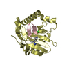
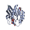
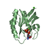
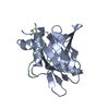
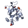
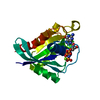
| Title | Cell attachment protein VP8* of a human rotavirus specifically interacts with A-type histo-blood group antigen | |||||||||
 Components Components | Outer capsid protein VP4 | |||||||||
 Keywords Keywords |  VIRAL PROTEIN / OTAVIRUS / CELL ATTACHMENT FACTOR / HISTO BLOOD GROUP ANTIGEN / GALECTIN-FOLD VIRAL PROTEIN / OTAVIRUS / CELL ATTACHMENT FACTOR / HISTO BLOOD GROUP ANTIGEN / GALECTIN-FOLD | |||||||||
| Function / homology |  Function and homology information Function and homology informationhost cell rough endoplasmic reticulum / host cytoskeleton / viral outer capsid / permeabilization of host organelle membrane involved in viral entry into host cell / symbiont entry into host cell via permeabilization of inner membrane / host cell endoplasmic reticulum-Golgi intermediate compartment / virion attachment to host cell / host cell plasma membrane / membraneSimilarity search - Function | |||||||||
| Biological species |  Rotavirus sp. Rotavirus sp. | |||||||||
| Method | X-RAY DIFFRACTION / MOLECULAR REPLACEMENT / Resolution: 1.56 Å | |||||||||
 Authors Authors | Hu, L. / Crawford, S.E. / Czako, R. / Cortes-Penfield, N.W. / Smith, D.F. / Le Pendu, J. / Estes, M.K. / Prasad, B.V.V. | |||||||||
 Citation Citation | Journal: Nature / Year: 2012 Title: Cell attachment protein VP8* of a human rotavirus specifically interacts with A-type histo-blood group antigen. Authors: Hu, L. / Crawford, S.E. / Czako, R. / Cortes-Penfield, N.W. / Smith, D.F. / Le Pendu, J. / Estes, M.K. / Prasad, B.V. | |||||||||
| History |
|
- Structure visualization
| Structure viewer | Molecule: MolmilJmol/JSmol |
|---|
- Downloads & links
-Download
| PDBx/mmCIF format | 4ds0.cif.gz | 87.5 KB | Display | PDBx/mmCIF format |
|---|---|---|---|---|
| PDB format | pdb4ds0.ent.gz | 62.5 KB | Display | PDB format |
| PDBx/mmJSON format | 4ds0.json.gz | Tree view | PDBx/mmJSON format | |
| Others |  Other downloads Other downloads |
-Validation report
| Arichive directory | https://data.pdbj.org/pub/pdb/validation_reports/ds/4ds0ftp://data.pdbj.org/pub/pdb/validation_reports/ds/4ds0 | HTTPS FTP |
|---|
-Related structure data
| Related structure data | 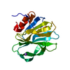 4drrC  4drvC  1kqrS C: citing same article ( S: Starting model for refinement |
|---|---|
| Similar structure data |


-Links
 PDBj
PDBj- Assembly
| Deposited unit | 
| ||||||||
|---|---|---|---|---|---|---|---|---|---|
| 1 |
| ||||||||
| Unit cell |
|
-Components
| #1: Protein | Mass: 18567.623 Da / Num. of mol.: 1 / Fragment: UNP residues 64-224 Source method: isolated from a genetically manipulated source Source: (gene. exp.) Rotavirus sp. / Gene: VP4 / Production host:  Escherichia coli (E. coli) / References: UniProt: Q86169 Escherichia coli (E. coli) / References: UniProt: Q86169 |
|---|---|
| #2: Polysaccharide | alpha-L-fucopyranose-(1-2)-[2-acetamido-2-deoxy-alpha-D-galactopyranose-(1-3)]beta-D- ...alpha-L-fucopyranose-(1-2)-[2-acetamido-2-deoxy-alpha-D-galactopyranose-(1-3)]beta-D-galactopyranose-(1-4)-2-acetamido-2-deoxy-beta-D-glucopyranose  , Oligosaccharide / Class: Antigen / Mass: 732.682 Da / Num. of mol.: 1 , Oligosaccharide / Class: Antigen / Mass: 732.682 Da / Num. of mol.: 1Source method: isolated from a genetically manipulated source Details: oligosaccharide with branches / References: Blood group A H type 2 antigen, beta anomer |
| #3: Water | ChemComp-HOH / Water Mass: 18.015 Da / Num. of mol.: 273 / Source method: isolated from a natural source / Formula: H2O Mass: 18.015 Da / Num. of mol.: 273 / Source method: isolated from a natural source / Formula: H2O |
-Experimental details
-Experiment
| Experiment | Method: X-RAY DIFFRACTION / Number of used crystals: 1 |
|---|
- Sample preparation
| Crystal | Density Matthews: 1.84 Å3/Da / Density % sol: 33.09 % |
|---|---|
| Crystal grow | Temperature: 293 K / Method: vapor diffusion, hanging drop / pH: 4.5 Details: 30% PEG 1500 Sodium acetate trihydrate, pH 4.5, VAPOR DIFFUSION, HANGING DROP, temperature 293K |
-Data collection
| Diffraction source | Source: ROTATING ANODE / Type: RIGAKU FR-E SUPERBRIGHT / Wavelength: 1.54 Å |
|---|---|
| Detector | Type: RIGAKU RAXIS HTC / Detector: IMAGE PLATE / Date: Nov 24, 2010 |
| Radiation | Protocol: SINGLE WAVELENGTH / Monochromatic (M) / Laue (L): M / Scattering type: x-ray |
| Radiation wavelength | Wavelength: 1.54 Å / Relative weight: 1 |
| Reflection | Resolution: 1.56→27.6 Å / Num. all: 19647 / Num. obs: 19647 / % possible obs: 92.6 % / Observed criterion σ(F): 2 / Observed criterion σ(I): 2 |
| Reflection shell | Resolution: 1.56→1.64 Å / % possible all: 86.8 |
- Processing
| Software | Name: PHENIX / Version: (phenix.refine: 1.4_162) / Classification: refinement | ||||||||||||||||||||||||||||||||||||||||||||||||||||||||||||||||||||||||||||||||||||||||||||||||||||
|---|---|---|---|---|---|---|---|---|---|---|---|---|---|---|---|---|---|---|---|---|---|---|---|---|---|---|---|---|---|---|---|---|---|---|---|---|---|---|---|---|---|---|---|---|---|---|---|---|---|---|---|---|---|---|---|---|---|---|---|---|---|---|---|---|---|---|---|---|---|---|---|---|---|---|---|---|---|---|---|---|---|---|---|---|---|---|---|---|---|---|---|---|---|---|---|---|---|---|---|---|---|
| Refinement | Method to determine structure: MOLECULAR REPLACEMENT Starting model: 1KQR Resolution: 1.56→27.6 Å / SU ML: 0.19 / σ(F): 1.35 / Phase error: 17.64 / Stereochemistry target values: ML
| ||||||||||||||||||||||||||||||||||||||||||||||||||||||||||||||||||||||||||||||||||||||||||||||||||||
| Solvent computation | Shrinkage radii: 0.9 Å / VDW probe radii: 1.11 Å / Solvent model: FLAT BULK SOLVENT MODEL / Bsol: 26.669 Å2 / ksol: 0.336 e/Å3 | ||||||||||||||||||||||||||||||||||||||||||||||||||||||||||||||||||||||||||||||||||||||||||||||||||||
| Displacement parameters |
| ||||||||||||||||||||||||||||||||||||||||||||||||||||||||||||||||||||||||||||||||||||||||||||||||||||
| Refinement step | Cycle: LAST / Resolution: 1.56→27.6 Å
| ||||||||||||||||||||||||||||||||||||||||||||||||||||||||||||||||||||||||||||||||||||||||||||||||||||
| Refine LS restraints |
| ||||||||||||||||||||||||||||||||||||||||||||||||||||||||||||||||||||||||||||||||||||||||||||||||||||
| LS refinement shell |
| ||||||||||||||||||||||||||||||||||||||||||||||||||||||||||||||||||||||||||||||||||||||||||||||||||||
| Refinement TLS params. | Method: refined / Refine-ID: X-RAY DIFFRACTION
| ||||||||||||||||||||||||||||||||||||||||||||||||||||||||||||||||||||||||||||||||||||||||||||||||||||
| Refinement TLS group |
|
