- PDB-4dqk: Crystal structure of the FAD binding domain of cytochrome P450 BM3 -
+
Open data
ID or keywords:
Loading...
-
Basic information
Entry
Database: PDB / ID: 4dqk
Title
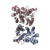
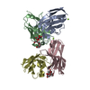
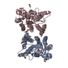
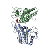
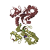
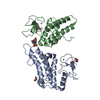
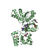
Crystal structure of the FAD binding domain of cytochrome P450 BM3
Components
Bifunctional P-450/NADPH-P450 reductase
Keywords
OXIDOREDUCTASE / Rossmann Fold / redox partner to cytochrome P450 / FAD and NADP binding / none
Function / homology
Function and homology information
NADPH-hemoprotein reductase / NADPH-hemoprotein reductase activity / oxidoreductase activity, acting on paired donors, with incorporation or reduction of molecular oxygen, reduced flavin or flavoprotein as one donor, and incorporation of one atom of oxygen / unspecific monooxygenase / aromatase activity / metabolic process / response to hormone / FMN binding / flavin adenine dinucleotide binding / iron ion binding ...NADPH-hemoprotein reductase / NADPH-hemoprotein reductase activity / oxidoreductase activity, acting on paired donors, with incorporation or reduction of molecular oxygen, reduced flavin or flavoprotein as one donor, and incorporation of one atom of oxygen / unspecific monooxygenase / aromatase activity / metabolic process / response to hormone / FMN binding / flavin adenine dinucleotide binding / iron ion binding / heme binding / identical protein binding / cytosol Similarity search - Function
Mass: 18.015 Da / Num. of mol.: 274 / Source method: isolated from a natural source / Formula: H2O
-
Experimental details
-
Experiment
Experiment
Method: X-RAY DIFFRACTION / Number of used crystals: 1
-
Sample preparation
Crystal
Density Matthews: 4.48 Å3/Da / Density % sol: 72.54 %
Crystal grow
Temperature: 293 K / Method: vapor diffusion, sitting drop / pH: 6.5 Details: 2 microL of mother liquor to 2 microL of 12 mg/ml FAD domain. Crystals were obtained using a well solution of 28% polyethylene glycol 8000, 0.3 M ammonium sulfate, cacodylate buffer pH 6.5. ...Details: 2 microL of mother liquor to 2 microL of 12 mg/ml FAD domain. Crystals were obtained using a well solution of 28% polyethylene glycol 8000, 0.3 M ammonium sulfate, cacodylate buffer pH 6.5. Crystals of dimensions 70 x 70 x 900 M formed after 4-7 days. In order to form a coenzyme complex with NADP+, C773A/C999A FAD domain crystals were soaked in a 10 mM NADP+ solution for 10 minutes., VAPOR DIFFUSION, SITTING DROP, temperature 293K
In the structure databanks used in Yorodumi, some data are registered as the other names, "COVID-19 virus" and "2019-nCoV". Here are the details of the virus and the list of structure data.
Jan 31, 2019. EMDB accession codes are about to change! (news from PDBe EMDB page)
EMDB accession codes are about to change! (news from PDBe EMDB page)
The allocation of 4 digits for EMDB accession codes will soon come to an end. Whilst these codes will remain in use, new EMDB accession codes will include an additional digit and will expand incrementally as the available range of codes is exhausted. The current 4-digit format prefixed with “EMD-” (i.e. EMD-XXXX) will advance to a 5-digit format (i.e. EMD-XXXXX), and so on. It is currently estimated that the 4-digit codes will be depleted around Spring 2019, at which point the 5-digit format will come into force.
The EM Navigator/Yorodumi systems omit the EMD- prefix.
Related info.:Q: What is EMD? / ID/Accession-code notation in Yorodumi/EM Navigator
Yorodumi is a browser for structure data from EMDB, PDB, SASBDB, etc.
This page is also the successor to EM Navigator detail page, and also detail information page/front-end page for Omokage search.
The word "yorodu" (or yorozu) is an old Japanese word meaning "ten thousand". "mi" (miru) is to see.
Related info.:EMDB / PDB / SASBDB / Comparison of 3 databanks / Yorodumi Search / Aug 31, 2016. New EM Navigator & Yorodumi / Yorodumi Papers / Jmol/JSmol / Function and homology information / Changes in new EM Navigator and Yorodumi
 Movie
Movie Controller

 Yorodumi
 Open data
 Basic information
 Components
 Keywords
 OXIDOREDUCTASE /
  Function and homology information
 Bacillus megaterium (bacteria)
 Authors
 Citation
 Structure visualization
 Downloads & links
 Other downloads
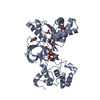


 PDBj
PDBj


 Assembly


 Mass: 785.550 Da / Num. of mol.: 2 / Source method: obtained synthetically / Formula: C27H33N9O15P2 / Comment: FAD*YM
 Mass: 96.063 Da / Num. of mol.: 6 / Source method: obtained synthetically / Formula: SO4
 Mass: 194.226 Da / Num. of mol.: 4 / Source method: obtained synthetically / Formula: C8H18O5 / Comment: precipitant*YM
 Mass: 18.015 Da / Num. of mol.: 274 / Source method: isolated from a natural source / Formula: H2O
 Sample preparation
 / Beamline: ID14-3 / Wavelength: 1 Å
 Processing