 Movie
Movie Controller

[English] 日本語
 Yorodumi
- PDB-4dnh: Crystal structure of hypothetical protein SMc04132 from Sinorhizo... -
+ Open data

- Basic information
| Entry | Database: PDB / ID: 4dnh | ||||||
|---|---|---|---|---|---|---|---|
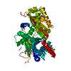
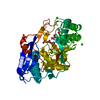
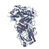
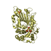
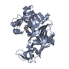
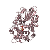
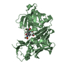
| Title | Crystal structure of hypothetical protein SMc04132 from Sinorhizobium meliloti 1021 | ||||||
 Components Components | Uncharacterized protein | ||||||
 Keywords Keywords |  Structural Genomics / Unknown Function / PSI-BIOLOGY / Protein Structure Initiative / New York Structural Genomics Research Consortium / NYSGRC Structural Genomics / Unknown Function / PSI-BIOLOGY / Protein Structure Initiative / New York Structural Genomics Research Consortium / NYSGRC | ||||||
| Function / homology | Protein of unknown function DUF993 / Protein of unknown function (DUF993) / Aldolase class I / TIM Barrel / Alpha-Beta Barrel / Alpha Beta / Uncharacterized protein Function and homology information Function and homology information | ||||||
| Biological species |  Sinorhizobium meliloti (bacteria) Sinorhizobium meliloti (bacteria) | ||||||
| Method | X-RAY DIFFRACTION / SYNCHROTRON / SAD / molecular replacement / Resolution: 2.5 Å | ||||||
 Authors Authors | Malashkevich, V.N. / Bhosle, R. / Toro, R. / Seidel, R. / Almo, S.C. / New York Structural Genomics Research Consortium (NYSGRC) | ||||||
 Citation Citation | Journal: To be Published Title: Crystal structure of hypothetical protein SMc04132 from Sinorhizobium meliloti 1021 Authors: Malashkevich, V.N. / Bhosle, R. / Toro, R. / Seidel, R. / Almo, S.C. | ||||||
| History |
|
- Structure visualization
| Structure viewer | Molecule: MolmilJmol/JSmol |
|---|
- Downloads & links
-Download
| PDBx/mmCIF format | 4dnh.cif.gz | 160.4 KB | Display | PDBx/mmCIF format |
|---|---|---|---|---|
| PDB format | pdb4dnh.ent.gz | 132.8 KB | Display | PDB format |
| PDBx/mmJSON format | 4dnh.json.gz | Tree view | PDBx/mmJSON format | |
| Others |  Other downloads Other downloads |
-Validation report
| Arichive directory | https://data.pdbj.org/pub/pdb/validation_reports/dn/4dnhftp://data.pdbj.org/pub/pdb/validation_reports/dn/4dnh | HTTPS FTP |
|---|
-Related structure data
| Similar structure data | |
|---|---|
| Other databases |


-Links
 PDBj
PDBj- Assembly
| Deposited unit | 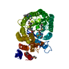
| ||||||||
|---|---|---|---|---|---|---|---|---|---|
| 1 |
| ||||||||
| Unit cell |
|
-Components
| #1: Protein | Mass: 43883.219 Da / Num. of mol.: 1 Source method: isolated from a genetically manipulated source Source: (gene. exp.) Sinorhizobium meliloti (bacteria) / Strain: 1021 / Gene: NP_384226.1, R00120, SMc04132 / Plasmid: BC-PSGX3(BC) / Production host: Escherichia coli (E. coli) / Strain (production host): BL21(DE3)CODON+RIL / References: UniProt: Q92T60 | ||
|---|---|---|---|
| #2: Chemical | Glycerol  Mass: 92.094 Da / Num. of mol.: 2 / Source method: obtained synthetically / Formula: C3H8O3 Mass: 92.094 Da / Num. of mol.: 2 / Source method: obtained synthetically / Formula: C3H8O3#3: Water | ChemComp-HOH / | Water Mass: 18.015 Da / Num. of mol.: 95 / Source method: isolated from a natural source / Formula: H2O Mass: 18.015 Da / Num. of mol.: 95 / Source method: isolated from a natural source / Formula: H2O |
-Experimental details
-Experiment
| Experiment | Method: X-RAY DIFFRACTION / Number of used crystals: 1 |
|---|
- Sample preparation
| Crystal | Density Matthews: 2.6 Å3/Da / Density % sol: 52.72 % / Mosaicity: 0.412 ° |
|---|---|
| Crystal grow | Temperature: 298 K / Method: vapor diffusion, sitting drop / pH: 7 Details: 20% PEG3000, 0.1 M Tris, pH 7.0, 0.2 M Ca(OAc), VAPOR DIFFUSION, SITTING DROP, temperature 298K |
-Data collection
| Diffraction | Mean temperature: 100 K | |||||||||||||||||||||||||||||||||||||||||||||||||||||||||||||||||||||||||||||||||||||||||||||||||||||||||||||||||||||||||||||||||||||||||||||||||||
|---|---|---|---|---|---|---|---|---|---|---|---|---|---|---|---|---|---|---|---|---|---|---|---|---|---|---|---|---|---|---|---|---|---|---|---|---|---|---|---|---|---|---|---|---|---|---|---|---|---|---|---|---|---|---|---|---|---|---|---|---|---|---|---|---|---|---|---|---|---|---|---|---|---|---|---|---|---|---|---|---|---|---|---|---|---|---|---|---|---|---|---|---|---|---|---|---|---|---|---|---|---|---|---|---|---|---|---|---|---|---|---|---|---|---|---|---|---|---|---|---|---|---|---|---|---|---|---|---|---|---|---|---|---|---|---|---|---|---|---|---|---|---|---|---|---|---|---|---|
| Diffraction source | Source: SYNCHROTRON / Site: NSLS  / Beamline: X29A / Wavelength: 0.9791 Å / Beamline: X29A / Wavelength: 0.9791 Å | |||||||||||||||||||||||||||||||||||||||||||||||||||||||||||||||||||||||||||||||||||||||||||||||||||||||||||||||||||||||||||||||||||||||||||||||||||
| Detector | Type: ADSC QUANTUM 315 / Detector: CCD / Date: Jun 17, 2009 | |||||||||||||||||||||||||||||||||||||||||||||||||||||||||||||||||||||||||||||||||||||||||||||||||||||||||||||||||||||||||||||||||||||||||||||||||||
| Radiation | Protocol: SINGLE WAVELENGTH / Scattering type: x-ray | |||||||||||||||||||||||||||||||||||||||||||||||||||||||||||||||||||||||||||||||||||||||||||||||||||||||||||||||||||||||||||||||||||||||||||||||||||
| Radiation wavelength | Wavelength: 0.9791 Å / Relative weight: 1 | |||||||||||||||||||||||||||||||||||||||||||||||||||||||||||||||||||||||||||||||||||||||||||||||||||||||||||||||||||||||||||||||||||||||||||||||||||
| Reflection | Redundancy: 3.8 % / Av σ(I) over netI: 13.64 / Number: 104452 / Rmerge(I) obs: 0.121 / Χ2: 1.33 / D res high: 2.6 Å / D res low: 50 Å / Num. obs: 27266 / % possible obs: 99.9 | |||||||||||||||||||||||||||||||||||||||||||||||||||||||||||||||||||||||||||||||||||||||||||||||||||||||||||||||||||||||||||||||||||||||||||||||||||
| Reflection | Resolution: 2.501→50 Å / Num. obs: 16435 / % possible obs: 99.9 % / Redundancy: 3.8 % / Rmerge(I) obs: 0.121 / Rrim(I) all: 0.121 / Χ2: 1.325 / Net I/av σ(I): 13.635 / Net I/σ(I): 7.1 / Num. measured all: 104452 | |||||||||||||||||||||||||||||||||||||||||||||||||||||||||||||||||||||||||||||||||||||||||||||||||||||||||||||||||||||||||||||||||||||||||||||||||||
| Reflection shell |
|
-Phasing
| Phasing | Method: molecular replacement | |||||||||
|---|---|---|---|---|---|---|---|---|---|---|
| Phasing MR | Rfactor: 53.99 / Model details: Phaser MODE: MR_AUTO
|
- Processing
| Software |
| |||||||||||||||||||||||||||||||||||||||||||||
|---|---|---|---|---|---|---|---|---|---|---|---|---|---|---|---|---|---|---|---|---|---|---|---|---|---|---|---|---|---|---|---|---|---|---|---|---|---|---|---|---|---|---|---|---|---|---|
| Refinement | Method to determine structure: SAD / Resolution: 2.5→20 Å / Cor.coef. Fo:Fc: 0.962 / Cor.coef. Fo:Fc free: 0.93 / WRfactor Rfree: 0.2113 / WRfactor Rwork: 0.1524 / Occupancy max: 1 / Occupancy min: 0.5 / FOM work R set: 0.8625 / SU B: 18.083 / SU ML: 0.198 / SU R Cruickshank DPI: 0.4285 / SU Rfree: 0.2715 / Cross valid method: THROUGHOUT / σ(F): 0 / ESU R: 0.428 / ESU R Free: 0.272 / Stereochemistry target values: MAXIMUM LIKELIHOOD Details: U VALUES : WITH TLS ADDED HYDROGENS HAVE BEEN USED IF PRESENT IN THE INPUT
| |||||||||||||||||||||||||||||||||||||||||||||
| Solvent computation | Ion probe radii: 0.8 Å / Shrinkage radii: 0.8 Å / VDW probe radii: 1.2 Å / Solvent model: MASK | |||||||||||||||||||||||||||||||||||||||||||||
| Displacement parameters | Biso max: 133.66 Å2 / Biso mean: 54.5714 Å2 / Biso min: 13.5 Å2
| |||||||||||||||||||||||||||||||||||||||||||||
| Refinement step | Cycle: LAST / Resolution: 2.5→20 Å
| |||||||||||||||||||||||||||||||||||||||||||||
| Refine LS restraints |
| |||||||||||||||||||||||||||||||||||||||||||||
| LS refinement shell | Resolution: 2.501→2.565 Å / Total num. of bins used: 20
| |||||||||||||||||||||||||||||||||||||||||||||
| Refinement TLS params. | Method: refined / Origin x: 40.5371 Å / Origin y: 7.986 Å / Origin z: 4.3952 Å
|
