 Movie
Movie Controller


+ Open data

- Basic information
| Entry | Database: PDB / ID: 4dnd | ||||||
|---|---|---|---|---|---|---|---|
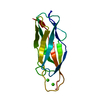
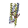
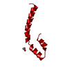
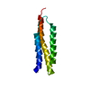
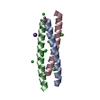
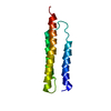
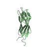
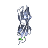
| Title | Crystal structure of syntaxin 10 from Homo sapiens | ||||||
 Components Components | Syntaxin-10 | ||||||
 Keywords Keywords | TRANSPORT PROTEIN / STRUCTURAL GENOMICS / PROTEIN STRUCTURE INITIATIVE / NYSGRC / PSI-Biology / New York Structural Genomics Research Consortium | ||||||
| Function / homology |  Function and homology information: / Retrograde transport at the Trans-Golgi-Network / Golgi vesicle transport / vesicle fusion / vesicle docking / SNARE complex / SNAP receptor activity / retrograde transport, endosome to Golgi / syntaxin binding / endomembrane system ...: / Retrograde transport at the Trans-Golgi-Network / Golgi vesicle transport / vesicle fusion / vesicle docking / SNARE complex / SNAP receptor activity / retrograde transport, endosome to Golgi / syntaxin binding / endomembrane system / SNARE binding / trans-Golgi network membrane / intracellular protein transport / trans-Golgi network / regulation of protein localization / synaptic vesicle / vesicle / membrane => GO:0016020 / perinuclear region of cytoplasm / cytosol Function and homology information: / Retrograde transport at the Trans-Golgi-Network / Golgi vesicle transport / vesicle fusion / vesicle docking / SNARE complex / SNAP receptor activity / retrograde transport, endosome to Golgi / syntaxin binding / endomembrane system ...: / Retrograde transport at the Trans-Golgi-Network / Golgi vesicle transport / vesicle fusion / vesicle docking / SNARE complex / SNAP receptor activity / retrograde transport, endosome to Golgi / syntaxin binding / endomembrane system / SNARE binding / trans-Golgi network membrane / intracellular protein transport / trans-Golgi network / regulation of protein localization / synaptic vesicle / vesicle / membrane => GO:0016020 / perinuclear region of cytoplasm / cytosolSimilarity search - Function | ||||||
| Biological species |  Homo sapiens (human) Homo sapiens (human) | ||||||
| Method | X-RAY DIFFRACTION / SYNCHROTRON / SAD / Resolution: 1.4 Å | ||||||
 Authors Authors | Malashkevich, V.N. / Bhosle, R. / Toro, R. / Seidel, R. / Almo, S.C. / New York Structural Genomics Research Consortium (NYSGRC) | ||||||
 Citation Citation | Journal: To be Published Title: Crystal structure of syntaxin 10 from Homo sapiens Authors: Malashkevich, V.N. / Bhosle, R. / Toro, R. / Seidel, R. / Almo, S.C. | ||||||
| History |
|
- Structure visualization
| Structure viewer | Molecule: MolmilJmol/JSmol |
|---|
- Downloads & links
-Download
| PDBx/mmCIF format | 4dnd.cif.gz | 55.5 KB | Display | PDBx/mmCIF format |
|---|---|---|---|---|
| PDB format | pdb4dnd.ent.gz | 42.4 KB | Display | PDB format |
| PDBx/mmJSON format | 4dnd.json.gz | Tree view | PDBx/mmJSON format | |
| Others |  Other downloads Other downloads |
-Validation report
| Arichive directory | https://data.pdbj.org/pub/pdb/validation_reports/dn/4dndftp://data.pdbj.org/pub/pdb/validation_reports/dn/4dnd | HTTPS FTP |
|---|
-Related structure data
| Similar structure data | |
|---|---|
| Other databases |


-Links
 PDBj
PDBj
- Assembly
| Deposited unit | 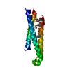
| ||||||||
|---|---|---|---|---|---|---|---|---|---|
| 1 |
| ||||||||
| Unit cell |
|
-Components
| #1: Protein | / Syn10 Mass: 15318.346 Da / Num. of mol.: 1 / Fragment: Cytoplasmic Topological domain residues 1-108 Source method: isolated from a genetically manipulated source Source: (gene. exp.) Homo sapiens (human) / Strain: Bronx / Gene: AAC05087.1, STX10, SYN10 / Plasmid: BC-PSGX3(BC) / Production host:  Escherichia coli (E. coli) / Strain (production host): BL21(DE3)CODON+RIL / References: UniProt: O60499 Escherichia coli (E. coli) / Strain (production host): BL21(DE3)CODON+RIL / References: UniProt: O60499 |
|---|---|
| #2: Water | ChemComp-HOH / Water Mass: 18.015 Da / Num. of mol.: 120 / Source method: isolated from a natural source / Formula: H2O Mass: 18.015 Da / Num. of mol.: 120 / Source method: isolated from a natural source / Formula: H2O |
-Experimental details
-Experiment
| Experiment | Method: X-RAY DIFFRACTION / Number of used crystals: 1 |
|---|
- Sample preparation
| Crystal | Density Matthews: 1.56 Å3/Da / Density % sol: 21.26 % |
|---|---|
| Crystal grow | Temperature: 298 K / Method: vapor diffusion, sitting drop / pH: 8.5 Details: 30% PEG400, 0.1M Tris pH 8.5, 0.2M MgCl2, VAPOR DIFFUSION, SITTING DROP, temperature 298K |
-Data collection
| Diffraction | Mean temperature: 100 K | |||||||||||||||||||||||||||||||||||||||||||||||||||||||||||||||||||||||||||||||||||||||||||||||||||||||||||||||||||||||||||||||||||||||||||||||||||
|---|---|---|---|---|---|---|---|---|---|---|---|---|---|---|---|---|---|---|---|---|---|---|---|---|---|---|---|---|---|---|---|---|---|---|---|---|---|---|---|---|---|---|---|---|---|---|---|---|---|---|---|---|---|---|---|---|---|---|---|---|---|---|---|---|---|---|---|---|---|---|---|---|---|---|---|---|---|---|---|---|---|---|---|---|---|---|---|---|---|---|---|---|---|---|---|---|---|---|---|---|---|---|---|---|---|---|---|---|---|---|---|---|---|---|---|---|---|---|---|---|---|---|---|---|---|---|---|---|---|---|---|---|---|---|---|---|---|---|---|---|---|---|---|---|---|---|---|---|
| Diffraction source | Source: SYNCHROTRON / Site: NSLS  / Beamline: X29A / Wavelength: 0.9791 Å / Beamline: X29A / Wavelength: 0.9791 Å | |||||||||||||||||||||||||||||||||||||||||||||||||||||||||||||||||||||||||||||||||||||||||||||||||||||||||||||||||||||||||||||||||||||||||||||||||||
| Detector | Type: ADSC QUANTUM 315 / Detector: CCD / Date: Dec 15, 2011 | |||||||||||||||||||||||||||||||||||||||||||||||||||||||||||||||||||||||||||||||||||||||||||||||||||||||||||||||||||||||||||||||||||||||||||||||||||
| Radiation | Protocol: SINGLE WAVELENGTH / Scattering type: x-ray | |||||||||||||||||||||||||||||||||||||||||||||||||||||||||||||||||||||||||||||||||||||||||||||||||||||||||||||||||||||||||||||||||||||||||||||||||||
| Radiation wavelength | Wavelength: 0.9791 Å / Relative weight: 1 | |||||||||||||||||||||||||||||||||||||||||||||||||||||||||||||||||||||||||||||||||||||||||||||||||||||||||||||||||||||||||||||||||||||||||||||||||||
| Reflection | Redundancy: 2.8 % / Av σ(I) over netI: 31.76 / Number: 95400 / Rmerge(I) obs: 0.046 / Χ2: 1.46 / D res high: 1.4 Å / D res low: 50 Å / Num. obs: 33753 / % possible obs: 94.1 | |||||||||||||||||||||||||||||||||||||||||||||||||||||||||||||||||||||||||||||||||||||||||||||||||||||||||||||||||||||||||||||||||||||||||||||||||||
| Diffraction reflection shell |
| |||||||||||||||||||||||||||||||||||||||||||||||||||||||||||||||||||||||||||||||||||||||||||||||||||||||||||||||||||||||||||||||||||||||||||||||||||
| Reflection | Resolution: 1.4→50 Å / Num. obs: 18149 / % possible obs: 94.1 % / Redundancy: 2.8 % / Rmerge(I) obs: 0.046 / Χ2: 1.46 / Net I/σ(I): 14.9 | |||||||||||||||||||||||||||||||||||||||||||||||||||||||||||||||||||||||||||||||||||||||||||||||||||||||||||||||||||||||||||||||||||||||||||||||||||
| Reflection shell |
|
-Phasing
| Phasing | Method: SAD |
|---|
- Processing
| Software |
| ||||||||||||||||||||||||||||||||||||||||||||||||||||||||||||
|---|---|---|---|---|---|---|---|---|---|---|---|---|---|---|---|---|---|---|---|---|---|---|---|---|---|---|---|---|---|---|---|---|---|---|---|---|---|---|---|---|---|---|---|---|---|---|---|---|---|---|---|---|---|---|---|---|---|---|---|---|---|
| Refinement | Method to determine structure: SAD / Resolution: 1.4→17.52 Å / Cor.coef. Fo:Fc: 0.965 / Cor.coef. Fo:Fc free: 0.955 / WRfactor Rfree: 0.2122 / WRfactor Rwork: 0.1692 / Occupancy max: 1 / Occupancy min: 0.5 / FOM work R set: 0.8781 / SU B: 2.4 / SU ML: 0.043 / SU R Cruickshank DPI: 0.0799 / SU Rfree: 0.0706 / Cross valid method: THROUGHOUT / σ(F): 0 / ESU R: 0.078 / ESU R Free: 0.068 / Stereochemistry target values: MAXIMUM LIKELIHOOD Details: HYDROGENS HAVE BEEN USED IF PRESENT IN THE INPUT U VALUES : RESIDUAL ONLY
| ||||||||||||||||||||||||||||||||||||||||||||||||||||||||||||
| Solvent computation | Ion probe radii: 0.8 Å / Shrinkage radii: 0.8 Å / VDW probe radii: 1.2 Å / Solvent model: MASK | ||||||||||||||||||||||||||||||||||||||||||||||||||||||||||||
| Displacement parameters | Biso max: 53.31 Å2 / Biso mean: 22.7954 Å2 / Biso min: 10.54 Å2
| ||||||||||||||||||||||||||||||||||||||||||||||||||||||||||||
| Refinement step | Cycle: LAST / Resolution: 1.4→17.52 Å
| ||||||||||||||||||||||||||||||||||||||||||||||||||||||||||||
| Refine LS restraints |
| ||||||||||||||||||||||||||||||||||||||||||||||||||||||||||||
| LS refinement shell | Resolution: 1.401→1.437 Å / Total num. of bins used: 20
| ||||||||||||||||||||||||||||||||||||||||||||||||||||||||||||
| Refinement TLS params. | Method: refined / Origin x: -0.1801 Å / Origin y: -0.641 Å / Origin z: 11.2748 Å
|
