 Movie
Movie Controller


+ Open data

- Basic information
| Entry | Database: PDB / ID: 4dnc | ||||||
|---|---|---|---|---|---|---|---|
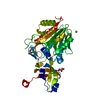
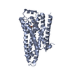
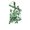
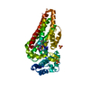
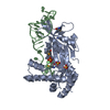
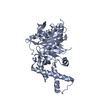
| Title | Crystal structure of human MOF in complex with MSL1 | ||||||
 Components Components |
| ||||||
 Keywords Keywords |  TRANSCRIPTION / histone acetyltransferase / MOF / MSL / NSL TRANSCRIPTION / histone acetyltransferase / MOF / MSL / NSL | ||||||
| Function / homology |  Function and homology information Function and homology informationMSL complex / histone H4K16 acetyltransferase activity / : / regulation of mRNA processing / myeloid cell differentiation / NSL complex / peptide-lysine-N-acetyltransferase activity / Formation of WDR5-containing histone-modifying complexes / MLL1 complex / histone acetyltransferase complex ...MSL complex / histone H4K16 acetyltransferase activity / : / regulation of mRNA processing / myeloid cell differentiation / NSL complex / peptide-lysine-N-acetyltransferase activity / Formation of WDR5-containing histone-modifying complexes / MLL1 complex / histone acetyltransferase complex / histone acetyltransferase / transcription initiation-coupled chromatin remodeling / neurogenesis / regulation of autophagy / transcription coregulator activity / kinetochore / nuclear matrix / HATs acetylate histones / RNA polymerase II-specific DNA-binding transcription factor binding / transcription coactivator activity / nuclear speck / chromatin remodeling / negative regulation of DNA-templated transcription / chromatin binding / positive regulation of DNA-templated transcription / enzyme binding / positive regulation of transcription by RNA polymerase II / nucleoplasm / metal ion binding / nucleusSimilarity search - Function | ||||||
| Biological species |  Homo sapiens (human) Homo sapiens (human) | ||||||
| Method | X-RAY DIFFRACTION / SYNCHROTRON / MOLECULAR REPLACEMENT / Resolution: 2.05 Å | ||||||
 Authors Authors | Huang, J. / Lei, M. | ||||||
 Citation Citation | Journal: Cell Res. / Year: 2012 Title: Structural insight into the regulation of MOF in the male-specific lethal complex and the non-specific lethal complex. Authors: Huang, J. / Wan, B. / Wu, L. / Yang, Y. / Dou, Y. / Lei, M. | ||||||
| History |
|
- Structure visualization
| Structure viewer | Molecule: MolmilJmol/JSmol |
|---|
- Downloads & links
-Download
| PDBx/mmCIF format | 4dnc.cif.gz | 139.5 KB | Display | PDBx/mmCIF format |
|---|---|---|---|---|
| PDB format | pdb4dnc.ent.gz | 114.7 KB | Display | PDB format |
| PDBx/mmJSON format | 4dnc.json.gz | Tree view | PDBx/mmJSON format | |
| Others |  Other downloads Other downloads |
-Validation report
| Arichive directory | https://data.pdbj.org/pub/pdb/validation_reports/dn/4dncftp://data.pdbj.org/pub/pdb/validation_reports/dn/4dnc | HTTPS FTP |
|---|
-Related structure data
| Similar structure data |
|---|


-Links
 PDBj
PDBj


- Assembly
| Deposited unit | 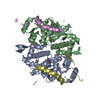
| ||||||||
|---|---|---|---|---|---|---|---|---|---|
| 1 | 
| ||||||||
| 2 | 
| ||||||||
| 3 |
| ||||||||
| Unit cell |
|
-Components
| #1: Protein | Mass: 33983.344 Da / Num. of mol.: 2 / Fragment: HAT domain (UNP residues 170-458) Source method: isolated from a genetically manipulated source Source: (gene. exp.) Homo sapiens (human) / Gene: KAT8, MOF, MYST1, PP7073 / Production host:  Escherichia coli (E. coli) / Strain (production host): BL21(DE3) / References: UniProt: Q9H7Z6, histone acetyltransferase Escherichia coli (E. coli) / Strain (production host): BL21(DE3) / References: UniProt: Q9H7Z6, histone acetyltransferase#2: Protein/peptide | Mass: 5764.471 Da / Num. of mol.: 2 / Fragment: MOF-binding motif (UNP residues 473-520) Source method: isolated from a genetically manipulated source Source: (gene. exp.) Homo sapiens (human) / Gene: MSL1, MSL1L1 / Production host: Escherichia coli (E. coli) / Strain (production host): BL21(DE3) / References: UniProt: Q68DK7#3: Chemical |   Mass: 65.409 Da / Num. of mol.: 2 / Source method: obtained synthetically / Formula: Zn Mass: 65.409 Da / Num. of mol.: 2 / Source method: obtained synthetically / Formula: Zn#4: Water | ChemComp-HOH / | Water Mass: 18.015 Da / Num. of mol.: 151 / Source method: isolated from a natural source / Formula: H2O Mass: 18.015 Da / Num. of mol.: 151 / Source method: isolated from a natural source / Formula: H2O |
|---|
-Experimental details
-Experiment
| Experiment | Method: X-RAY DIFFRACTION / Number of used crystals: 1 |
|---|
- Sample preparation
| Crystal | Density Matthews: 2.83 Å3/Da / Density % sol: 56.56 % |
|---|---|
| Crystal grow | Temperature: 277 K / Method: vapor diffusion, sitting drop / pH: 7 Details: 1.6 M Li2SO4, pH 7.0, VAPOR DIFFUSION, SITTING DROP, temperature 277K |
-Data collection
| Diffraction | Mean temperature: 100 K |
|---|---|
| Diffraction source | Source: SYNCHROTRON / Site: APS  / Beamline: 21-ID-D / Wavelength: 0.98159 Å / Beamline: 21-ID-D / Wavelength: 0.98159 Å |
| Detector | Type: MARMOSAIC 300 mm CCD / Detector: CCD / Date: Mar 20, 2010 |
| Radiation | Protocol: SINGLE WAVELENGTH / Monochromatic (M) / Laue (L): M / Scattering type: x-ray |
| Radiation wavelength | Wavelength: 0.98159 Å / Relative weight: 1 |
| Reflection | Resolution: 2.05→100 Å / Num. obs: 54362 |
- Processing
| Software |
| |||||||||||||||||||||||||||||||||||||||||||||||||||||||||||||||||||||||||||||||||||||||||||||||||||||||||||||||||||||||||||||||||||||||||||||||||||||||||||||||||||||||||||||||||||||||||||||||||||||||||||||||||||||||||
|---|---|---|---|---|---|---|---|---|---|---|---|---|---|---|---|---|---|---|---|---|---|---|---|---|---|---|---|---|---|---|---|---|---|---|---|---|---|---|---|---|---|---|---|---|---|---|---|---|---|---|---|---|---|---|---|---|---|---|---|---|---|---|---|---|---|---|---|---|---|---|---|---|---|---|---|---|---|---|---|---|---|---|---|---|---|---|---|---|---|---|---|---|---|---|---|---|---|---|---|---|---|---|---|---|---|---|---|---|---|---|---|---|---|---|---|---|---|---|---|---|---|---|---|---|---|---|---|---|---|---|---|---|---|---|---|---|---|---|---|---|---|---|---|---|---|---|---|---|---|---|---|---|---|---|---|---|---|---|---|---|---|---|---|---|---|---|---|---|---|---|---|---|---|---|---|---|---|---|---|---|---|---|---|---|---|---|---|---|---|---|---|---|---|---|---|---|---|---|---|---|---|---|---|---|---|---|---|---|---|---|---|---|---|---|---|---|---|---|
| Refinement | Method to determine structure: MOLECULAR REPLACEMENT / Resolution: 2.05→40.994 Å / SU ML: 0.28 / σ(F): 1.34 / Phase error: 26.3 / Stereochemistry target values: ML
| |||||||||||||||||||||||||||||||||||||||||||||||||||||||||||||||||||||||||||||||||||||||||||||||||||||||||||||||||||||||||||||||||||||||||||||||||||||||||||||||||||||||||||||||||||||||||||||||||||||||||||||||||||||||||
| Solvent computation | Shrinkage radii: 0.83 Å / VDW probe radii: 1.1 Å / Solvent model: FLAT BULK SOLVENT MODEL / Bsol: 43.572 Å2 / ksol: 0.388 e/Å3 | |||||||||||||||||||||||||||||||||||||||||||||||||||||||||||||||||||||||||||||||||||||||||||||||||||||||||||||||||||||||||||||||||||||||||||||||||||||||||||||||||||||||||||||||||||||||||||||||||||||||||||||||||||||||||
| Displacement parameters |
| |||||||||||||||||||||||||||||||||||||||||||||||||||||||||||||||||||||||||||||||||||||||||||||||||||||||||||||||||||||||||||||||||||||||||||||||||||||||||||||||||||||||||||||||||||||||||||||||||||||||||||||||||||||||||
| Refinement step | Cycle: LAST / Resolution: 2.05→40.994 Å
| |||||||||||||||||||||||||||||||||||||||||||||||||||||||||||||||||||||||||||||||||||||||||||||||||||||||||||||||||||||||||||||||||||||||||||||||||||||||||||||||||||||||||||||||||||||||||||||||||||||||||||||||||||||||||
| Refine LS restraints |
| |||||||||||||||||||||||||||||||||||||||||||||||||||||||||||||||||||||||||||||||||||||||||||||||||||||||||||||||||||||||||||||||||||||||||||||||||||||||||||||||||||||||||||||||||||||||||||||||||||||||||||||||||||||||||
| LS refinement shell |
|
