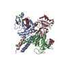
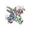
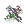
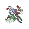
Toll Like Receptor 4 (TLR4) Cascade / Signal regulatory protein family interactions / Regulation of TLR by endogenous ligand / Surfactant metabolism / Immunoregulatory interactions between a Lymphoid and a non-Lymphoid cell / extracellular matrix structural constituent conferring tensile strength / respiratory gaseous exchange by respiratory system / collagen trimer / surfactant homeostasis / lung alveolus development ...Toll Like Receptor 4 (TLR4) Cascade / Signal regulatory protein family interactions / Regulation of TLR by endogenous ligand / Surfactant metabolism / Immunoregulatory interactions between a Lymphoid and a non-Lymphoid cell / extracellular matrix structural constituent conferring tensile strength / respiratory gaseous exchange by respiratory system / collagen trimer / surfactant homeostasis / lung alveolus development / positive regulation of phagocytosis / multivesicular body / extracellular matrix organization / carbohydrate binding / collagen-containing extracellular matrix / innate immune response / extracellular space Similarity search - Function
Lung surfactant protein D coiled-coil trimerisation / Lung surfactant protein D coiled-coil trimerisation / Collagen triple helix repeat / Collagen triple helix repeat (20 copies) / C-type lectin, conserved site / C-type lectin domain signature. / Mannose-Binding Protein A; Chain A / Mannose-Binding Protein A, subunit A / Lectin C-type domain / C-type lectin domain profile. ...Lung surfactant protein D coiled-coil trimerisation / Lung surfactant protein D coiled-coil trimerisation / Collagen triple helix repeat / Collagen triple helix repeat (20 copies) / C-type lectin, conserved site / C-type lectin domain signature. / Mannose-Binding Protein A; Chain A / Mannose-Binding Protein A, subunit A / Lectin C-type domain / C-type lectin domain profile. / C-type lectin-like / C-type lectin (CTL) or carbohydrate-recognition domain (CRD) / C-type lectin-like/link domain superfamily / C-type lectin fold / Roll / Alpha Beta Similarity search - Domain/homology
Mass: 18.015 Da / Num. of mol.: 36 / Source method: isolated from a natural source / Formula: H2O
-
Experimental details
-
Experiment
Experiment
Method: X-RAY DIFFRACTION / Number of used crystals: 1
-
Sample preparation
Crystal
Density Matthews: 2.44 Å3/Da / Density % sol: 49.49 %
Crystal grow
Temperature: 290 K / Method: vapor diffusion, hanging drop / pH: 6.5 Details: 12.3 mg/ml protein in 0.9 % sodium chloride, 5 mM EDTA, 15 mM calcium chloride, and 20 mM mannose was mixed with an equal volume of 0.1 M bistris (pH =6.5), 20% w/v PEG 3350, 200 mM magesium ...Details: 12.3 mg/ml protein in 0.9 % sodium chloride, 5 mM EDTA, 15 mM calcium chloride, and 20 mM mannose was mixed with an equal volume of 0.1 M bistris (pH =6.5), 20% w/v PEG 3350, 200 mM magesium chloride, and 15% v/v glycerol. Prior to data collection crystals were soaked in a 30% w/v D-mannose solution., VAPOR DIFFUSION, HANGING DROP, temperature 290K
Resolution: 2.2→2.28 Å / Redundancy: 9.4 % / Rmerge(I) obs: 0.418 / Mean I/σ(I) obs: 7.25 / Num. unique all: 839 / % possible all: 99.3
-
Processing
Software
Name
Version
Classification
CrystalClear
datacollection
PHASER
phasing
PHENIX
(phenix.refine: 1.6.3_473)
refinement
DENZO
datareduction
SCALEPACK
datascaling
Refinement
Method to determine structure: FOURIER SYNTHESIS Starting model: A complex of pig SPD and glycerol solved by molecular replacement in the same space group using human SPD (pdb code 2GGU B chain)
In the structure databanks used in Yorodumi, some data are registered as the other names, "COVID-19 virus" and "2019-nCoV". Here are the details of the virus and the list of structure data.
Jan 31, 2019. EMDB accession codes are about to change! (news from PDBe EMDB page)
EMDB accession codes are about to change! (news from PDBe EMDB page)
The allocation of 4 digits for EMDB accession codes will soon come to an end. Whilst these codes will remain in use, new EMDB accession codes will include an additional digit and will expand incrementally as the available range of codes is exhausted. The current 4-digit format prefixed with “EMD-” (i.e. EMD-XXXX) will advance to a 5-digit format (i.e. EMD-XXXXX), and so on. It is currently estimated that the 4-digit codes will be depleted around Spring 2019, at which point the 5-digit format will come into force.
The EM Navigator/Yorodumi systems omit the EMD- prefix.
Related info.:Q: What is EMD? / ID/Accession-code notation in Yorodumi/EM Navigator
Yorodumi is a browser for structure data from EMDB, PDB, SASBDB, etc.
This page is also the successor to EM Navigator detail page, and also detail information page/front-end page for Omokage search.
The word "yorodu" (or yorozu) is an old Japanese word meaning "ten thousand". "mi" (miru) is to see.
Related info.:EMDB / PDB / SASBDB / Comparison of 3 databanks / Yorodumi Search / Aug 31, 2016. New EM Navigator & Yorodumi / Yorodumi Papers / Jmol/JSmol / Function and homology information / Changes in new EM Navigator and Yorodumi
 Movie
Movie Controller

 Yorodumi
 Open data
 Basic information
 Components
 Keywords
 collectin /
  Function and homology information
 Authors
 Citation
 Structure visualization
 Downloads & links
 Other downloads

 PDBj
PDBj


 Assembly


 Mass: 40.078 Da / Num. of mol.: 2 / Source method: obtained synthetically / Formula: Ca
 Type: D-saccharide, beta linking / Mass: 180.156 Da / Num. of mol.: 1
 Mass: 18.015 Da / Num. of mol.: 36 / Source method: isolated from a natural source / Formula: H2O
 Sample preparation
 Processing