 Movie
Movie Controller

[English] 日本語
 Yorodumi
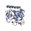
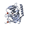
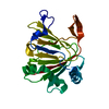
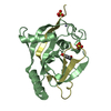
- PDB-4dlh: Crystal Structure of the protein Q9HRE7 from Halobacterium salina... -
+ Open data

- Basic information
| Entry | Database: PDB / ID: 4dlh | ||||||
|---|---|---|---|---|---|---|---|
| Title | Crystal Structure of the protein Q9HRE7 from Halobacterium salinarium at the resolution 1.9A, Northeast Structural Genomics Consortium (NESG) Target HsR50 | ||||||
 Components Components | uncharacterized protein | ||||||
 Keywords Keywords |  Structural Genomics / Unknown Function / PSI-Biology / Protein Structure Initiative / Northeast Structural Genomics Consortium (NESG) Structural Genomics / Unknown Function / PSI-Biology / Protein Structure Initiative / Northeast Structural Genomics Consortium (NESG) | ||||||
| Function / homology | Protein of unknown function DUF1684 / Protein of unknown function (DUF1684) / DUF1684 family protein Function and homology information Function and homology information | ||||||
| Biological species |  Halobacterium sp. NRC-1 (Halophile) Halobacterium sp. NRC-1 (Halophile) | ||||||
| Method | X-RAY DIFFRACTION / SYNCHROTRON / MOLECULAR REPLACEMENT / Resolution: 1.9 Å | ||||||
 Authors Authors | Kuzin, A. / Chen, Y. / Seetharaman, J. / Mao, M. / Xiao, R. / Ciccosanti, C. / Zhao, L. / Everett, J.K. / Nair, R. / Acton, T.B. ...Kuzin, A. / Chen, Y. / Seetharaman, J. / Mao, M. / Xiao, R. / Ciccosanti, C. / Zhao, L. / Everett, J.K. / Nair, R. / Acton, T.B. / Rost, B. / Montelione, G.T. / Hunt, J.F. / Tong, L. / Northeast Structural Genomics Consortium (NESG) | ||||||
 Citation Citation | Journal: To be Published Title: Northeast Structural Genomics Consortium Target HsR50 Authors: Kuzin, A. / Chen, Y. / Seetharaman, J. / Mao, M. / Xiao, R. / Ciccosanti, C. / Zhao, L. / Everett, J.K. / Nair, R. / Acton, T.B. / Rost, B. / Montelione, G.T. / Hunt, J.F. / Tong, L. | ||||||
| History |
|
- Structure visualization
| Structure viewer | Molecule: MolmilJmol/JSmol |
|---|
- Downloads & links
-Download
| PDBx/mmCIF format | 4dlh.cif.gz | 158.8 KB | Display | PDBx/mmCIF format |
|---|---|---|---|---|
| PDB format | pdb4dlh.ent.gz | 126.9 KB | Display | PDB format |
| PDBx/mmJSON format | 4dlh.json.gz | Tree view | PDBx/mmJSON format | |
| Others |  Other downloads Other downloads |
-Validation report
| Arichive directory | https://data.pdbj.org/pub/pdb/validation_reports/dl/4dlhftp://data.pdbj.org/pub/pdb/validation_reports/dl/4dlh | HTTPS FTP |
|---|
-Related structure data
| Related structure data |  4fj4C 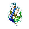 2lokS S: Starting model for refinement C: citing same article ( |
|---|---|
| Similar structure data | |
| Other databases |


-Links
 PDBj
PDBj- Assembly
| Deposited unit | 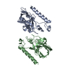
| ||||||||
|---|---|---|---|---|---|---|---|---|---|
| 1 | 
| ||||||||
| 2 | 
| ||||||||
| Unit cell |
| ||||||||
| Details | monomer,23.9 kD,91.6% |
-Components
| #1: Protein | Mass: 21582.410 Da / Num. of mol.: 2 Source method: isolated from a genetically manipulated source Source: (gene. exp.) Halobacterium sp. NRC-1 (Halophile) / References: UniProt: Q9HRE7#2: Water | ChemComp-HOH / | Water Mass: 18.015 Da / Num. of mol.: 337 / Source method: isolated from a natural source / Formula: H2O Mass: 18.015 Da / Num. of mol.: 337 / Source method: isolated from a natural source / Formula: H2O |
|---|
-Experimental details
-Experiment
| Experiment | Method: X-RAY DIFFRACTION / Number of used crystals: 1 |
|---|
- Sample preparation
| Crystal | Density Matthews: 2.18 Å3/Da / Density % sol: 43.6 % |
|---|---|
| Crystal grow | Temperature: 293 K / Method: mocrobatch under oil / pH: 7.5 Details: Protein solution: 100mM NaCl, 5mM DTT, 0.02% NaN3, 10mM Tris-HCl (pH 7.5) . Reservoir solution:KH2PO4 0.1M, HEPES 0.1M, PEG 8000 40%., mocrobatch under oil, temperature 293KK |
-Data collection
| Diffraction | Mean temperature: 100 K |
|---|---|
| Diffraction source | Source: SYNCHROTRON / Site: NSLS  / Beamline: X4A / Wavelength: 0.979 Å / Beamline: X4A / Wavelength: 0.979 Å |
| Detector | Type: ADSC QUANTUM 4 / Detector: CCD / Date: Apr 21, 2008 / Details: mirrors |
| Radiation | Monochromator: Si 111 CHANNEL / Protocol: SINGLE WAVELENGTH / Monochromatic (M) / Laue (L): M / Scattering type: x-ray |
| Radiation wavelength | Wavelength: 0.979 Å / Relative weight: 1 |
| Reflection | Resolution: 1.9→50 Å / Num. obs: 54970 / % possible obs: 95.5 % / Observed criterion σ(I): -3 / Redundancy: 3 % / Biso Wilson estimate: 22.33 Å2 / Rmerge(I) obs: 0.056 / Net I/σ(I): 23.7 |
| Reflection shell | Resolution: 1.9→1.97 Å / Redundancy: 2.5 % / Rmerge(I) obs: 0.172 / % possible all: 72.5 |
- Processing
| Software |
| ||||||||||||||||||||||||||||||||||||||||||||||||||||||||||||||||||||||||||||||||||||
|---|---|---|---|---|---|---|---|---|---|---|---|---|---|---|---|---|---|---|---|---|---|---|---|---|---|---|---|---|---|---|---|---|---|---|---|---|---|---|---|---|---|---|---|---|---|---|---|---|---|---|---|---|---|---|---|---|---|---|---|---|---|---|---|---|---|---|---|---|---|---|---|---|---|---|---|---|---|---|---|---|---|---|---|---|---|
| Refinement | Method to determine structure: MOLECULAR REPLACEMENT Starting model: pdb entry 2LOK Resolution: 1.9→38.244 Å / Occupancy max: 1 / Occupancy min: 0.5 / FOM work R set: 0.856 / SU ML: 0.19 / Cross valid method: throughput / σ(F): 1.34 / Phase error: 21.07 / Stereochemistry target values: ML
| ||||||||||||||||||||||||||||||||||||||||||||||||||||||||||||||||||||||||||||||||||||
| Solvent computation | Shrinkage radii: 0.6 Å / VDW probe radii: 0.9 Å / Solvent model: FLAT BULK SOLVENT MODEL / Bsol: 41.96 Å2 / ksol: 0.378 e/Å3 | ||||||||||||||||||||||||||||||||||||||||||||||||||||||||||||||||||||||||||||||||||||
| Displacement parameters | Biso max: 92.83 Å2 / Biso mean: 29.543 Å2 / Biso min: 8.6 Å2
| ||||||||||||||||||||||||||||||||||||||||||||||||||||||||||||||||||||||||||||||||||||
| Refinement step | Cycle: LAST / Resolution: 1.9→38.244 Å
| ||||||||||||||||||||||||||||||||||||||||||||||||||||||||||||||||||||||||||||||||||||
| Refine LS restraints |
| ||||||||||||||||||||||||||||||||||||||||||||||||||||||||||||||||||||||||||||||||||||
| LS refinement shell | Refine-ID: X-RAY DIFFRACTION / Total num. of bins used: 11
| ||||||||||||||||||||||||||||||||||||||||||||||||||||||||||||||||||||||||||||||||||||
| Refinement TLS params. | Method: refined / Origin x: 25.1288 Å / Origin y: 38.7723 Å / Origin z: 67.8398 Å
| ||||||||||||||||||||||||||||||||||||||||||||||||||||||||||||||||||||||||||||||||||||
| Refinement TLS group |
|
