 Movie
Movie Controller

[English] 日本語
 Yorodumi
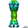
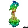
- PDB-4dk0: Crystal structure of MacA from Actinobacillus actinomycetemcomitans -
+ Open data

- Basic information
| Entry | Database: PDB / ID: 4dk0 | ||||||
|---|---|---|---|---|---|---|---|
| Title | Crystal structure of MacA from Actinobacillus actinomycetemcomitans | ||||||
 Components Components | Putative MacA | ||||||
 Keywords Keywords |  MEMBRANE PROTEIN / alpha-hairpin / lipoyl / beta-barrel / PERIPLASMIC PROTEIN MEMBRANE PROTEIN / alpha-hairpin / lipoyl / beta-barrel / PERIPLASMIC PROTEIN | ||||||
| Function / homology |  Function and homology information Function and homology informationmacrolide transmembrane transporter complex / xenobiotic detoxification by transmembrane export across the plasma membrane / extrinsic component of membrane / transmembrane transporter activity / identical protein bindingSimilarity search - Function | ||||||
| Biological species |  Aggregatibacter actinomycetemcomitans (bacteria) Aggregatibacter actinomycetemcomitans (bacteria) | ||||||
| Method | X-RAY DIFFRACTION / SYNCHROTRON / MAD / Resolution: 3.5 Å | ||||||
 Authors Authors | Xu, Y. / Piao, S. / Ha, N.C. | ||||||
 Citation Citation | Journal: J Biol Chem / Year: 2012 Title: Assembly and channel opening of outer membrane protein in tripartite drug efflux pumps of Gram-negative bacteria. Authors: Yongbin Xu / Arne Moeller / So-Young Jun / Minho Le / Bo-Young Yoon / Jin-Sik Kim / Kangseok Lee / Nam-Chul Ha /  Abstract: Gram-negative bacteria are capable of expelling diverse xenobiotic substances from within the cell by use of three-component efflux pumps in which the energy-activated inner membrane transporter is ...Gram-negative bacteria are capable of expelling diverse xenobiotic substances from within the cell by use of three-component efflux pumps in which the energy-activated inner membrane transporter is connected to the outer membrane channel protein via the membrane fusion protein. In this work, we describe the crystal structure of the membrane fusion protein MexA from the Pseudomonas aeruginosa MexAB-OprM pump in the hexameric ring arrangement. Electron microscopy study on the chimeric complex of MexA and the outer membrane protein OprM reveals that MexA makes a tip-to-tip interaction with OprM, which suggests a docking model for MexA and OprM. This docking model agrees well with genetic results and depicts detailed interactions. Opening of the OprM channel is accompanied by the simultaneous exposure of a protein structure resembling a six-bladed cogwheel, which intermeshes with the complementary cogwheel structure in the MexA hexamer. Taken together, we suggest an assembly and channel opening model for the MexAB-OprM pump. This study provides a better understanding of multidrug resistance in Gram-negative bacteria. | ||||||
| History |
|
- Structure visualization
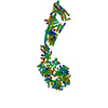
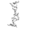
| Structure viewer | Molecule: MolmilJmol/JSmol |
|---|
- Downloads & links
-Download
| PDBx/mmCIF format | 4dk0.cif.gz | 138.6 KB | Display | PDBx/mmCIF format |
|---|---|---|---|---|
| PDB format | pdb4dk0.ent.gz | 115.5 KB | Display | PDB format |
| PDBx/mmJSON format | 4dk0.json.gz | Tree view | PDBx/mmJSON format | |
| Others |  Other downloads Other downloads |
-Validation report
| Arichive directory | https://data.pdbj.org/pub/pdb/validation_reports/dk/4dk0ftp://data.pdbj.org/pub/pdb/validation_reports/dk/4dk0 | HTTPS FTP |
|---|
-Related structure data


-Links
 PDBj
PDBj
- Assembly
| Deposited unit | 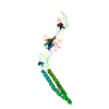
| ||||||||
|---|---|---|---|---|---|---|---|---|---|
| 1 | x 12
| ||||||||
| 2 | x 6
| ||||||||
| Unit cell |
|
-Components
| #1: Protein | Mass: 39866.645 Da / Num. of mol.: 1 / Fragment: UNP RESIDUES 30-394 Source method: isolated from a genetically manipulated source Source: (gene. exp.) Aggregatibacter actinomycetemcomitans (bacteria)Gene: macA / Production host: Escherichia coli (E. coli) / References: UniProt: Q2EHL9 |
|---|---|
| Sequence details | THESE RESIDUES DERIVE FROM A VARIANT OF THE STRAIN OF THE BACTERIA |
-Experimental details
-Experiment
| Experiment | Method: X-RAY DIFFRACTION / Number of used crystals: 1 |
|---|
- Sample preparation
| Crystal | Density Matthews: 5.51 Å3/Da / Density % sol: 77.69 % |
|---|---|
| Crystal grow | Temperature: 277 K / Method: evaporation / pH: 8.5 Details: 0.01M Nickel chloride, 0.1M Tris-HCl, 1M Lithium Sulfate, pH 8.5, EVAPORATION, temperature 277K |
-Data collection
| Diffraction | Mean temperature: 100 K | |||||||||||||||
|---|---|---|---|---|---|---|---|---|---|---|---|---|---|---|---|---|
| Diffraction source | Source: SYNCHROTRON / Site: PAL/PLS / Beamline: 6C1 / Wavelength: 1.0000, 0.9794, 0.9796, 1.1271 | |||||||||||||||
| Detector | Type: ADSC QUANTUM 210 / Detector: CCD / Date: Jan 15, 2008 | |||||||||||||||
| Radiation | Monochromator: Double mirror / Protocol: MAD / Monochromatic (M) / Laue (L): M / Scattering type: x-ray | |||||||||||||||
| Radiation wavelength |
| |||||||||||||||
| Reflection | Resolution: 3→50 Å / Num. all: 18344 / Num. obs: 17810 / % possible obs: 97.2 % / Observed criterion σ(F): 0 / Observed criterion σ(I): 0 | |||||||||||||||
| Reflection shell | Resolution: 3→3.11 Å / % possible all: 87.1 |
- Processing
| Software |
| |||||||||||||||||||||||||||||||||||||||||||||||||||||||||||||||
|---|---|---|---|---|---|---|---|---|---|---|---|---|---|---|---|---|---|---|---|---|---|---|---|---|---|---|---|---|---|---|---|---|---|---|---|---|---|---|---|---|---|---|---|---|---|---|---|---|---|---|---|---|---|---|---|---|---|---|---|---|---|---|---|---|
| Refinement | Method to determine structure: MAD / Resolution: 3.5→9.991 Å / SU ML: 1.08 / σ(F): 0.17 / Phase error: 40.2 / Stereochemistry target values: MLHL Details: THE AUTHOR DID NOT USE THE DATA IN 3.5-3.0A SINCE THE R-FACTOR WAS NOT REASONABLE AND THE MAP QUALITY LOOKED LIKE 3.5A RESOLUTION MAP.
| |||||||||||||||||||||||||||||||||||||||||||||||||||||||||||||||
| Solvent computation | Shrinkage radii: 0.98 Å / VDW probe radii: 1.2 Å / Solvent model: FLAT BULK SOLVENT MODEL / Bsol: 111.379 Å2 / ksol: 0.317 e/Å3 | |||||||||||||||||||||||||||||||||||||||||||||||||||||||||||||||
| Displacement parameters |
| |||||||||||||||||||||||||||||||||||||||||||||||||||||||||||||||
| Refinement step | Cycle: LAST / Resolution: 3.5→9.991 Å
| |||||||||||||||||||||||||||||||||||||||||||||||||||||||||||||||
| Refine LS restraints |
| |||||||||||||||||||||||||||||||||||||||||||||||||||||||||||||||
| LS refinement shell |
| |||||||||||||||||||||||||||||||||||||||||||||||||||||||||||||||
| Refinement TLS params. | Method: refined / Origin x: 80.678 Å / Origin y: 104.0272 Å / Origin z: 76.7551 Å
| |||||||||||||||||||||||||||||||||||||||||||||||||||||||||||||||
| Refinement TLS group | Selection details: all |
