- PDB-4ddw: Thermotoga maritima reverse gyrase, c-centered orthorhombic form -
+
Open data
ID or keywords:
Loading...
-
Basic information
Entry
Database: PDB / ID: 4ddw
Title
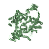
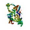
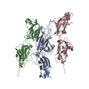
Thermotoga maritima reverse gyrase, c-centered orthorhombic form
Components
Reverse gyrase
Keywords
HYDROLASE / TOPOISOMERASE / DNA SUPERCOILING / ARCHAEA / HELICASE
Function / homology
Function and homology information
Isomerases; Isomerases altering macromolecular conformation; Enzymes altering nucleic acid conformation / reverse gyrase activity / DNA topoisomerase type II (double strand cut, ATP-hydrolyzing) activity / DNA unwinding involved in DNA replication / DNA topological change / helicase activity / ATP hydrolysis activity / DNA binding / zinc ion binding / ATP binding / cytoplasm Similarity search - Function
Phenylalanyl-tRNA Synthetase; Chain B, domain 1 - #80 / EV matrix protein fold - #20 / Reverse gyrase, zinc finger / Reverse gyrase zinc finger / EV matrix protein fold / Reverse gyrase / Reverse gyrase, TOPRIM domain / Zinc finger reverse gyrase N-terminal-type profile. / Zinc finger reverse gyrase C-terminal-type profile. / Topoisomerase I; domain 2 ...Phenylalanyl-tRNA Synthetase; Chain B, domain 1 - #80 / EV matrix protein fold - #20 / Reverse gyrase, zinc finger / Reverse gyrase zinc finger / EV matrix protein fold / Reverse gyrase / Reverse gyrase, TOPRIM domain / Zinc finger reverse gyrase N-terminal-type profile. / Zinc finger reverse gyrase C-terminal-type profile. / Topoisomerase I; domain 2 / Topoisomerase I, domain 2 / Rossmann fold - #140 / Topoisomerase I; domain 4 / Topoisomerase I, domain 4 / Topoisomerase (Topo) IA-type catalytic domain profile. / DNA topoisomerase, type IA, domain 2 / DNA topoisomerase, type IA, DNA-binding domain / DNA topoisomerase, type IA, central / DNA topoisomerase, type IA, central region, subdomain 1 / DNA topoisomerase, type IA, central region, subdomain 3 / DNA topoisomerase, type IA, core domain / DNA topoisomerase / Bacterial DNA topoisomeraes I ATP-binding domain / Bacterial DNA topoisomerase I DNA-binding domain / Phenylalanyl-tRNA Synthetase; Chain B, domain 1 / TOPRIM / Toprim domain / Toprim domain profile. / TOPRIM domain / DEAD/DEAH box helicase / DEAD/DEAH box helicase domain / Superfamilies 1 and 2 helicase C-terminal domain profile. / Superfamilies 1 and 2 helicase ATP-binding type-1 domain profile. / DEAD-like helicases superfamily / Helicase superfamily 1/2, ATP-binding domain / P-loop containing nucleotide triphosphate hydrolases / ATPases associated with a variety of cellular activities / AAA+ ATPase domain / Sandwich / P-loop containing nucleoside triphosphate hydrolase / Rossmann fold / 2-Layer Sandwich / Orthogonal Bundle / 3-Layer(aba) Sandwich / Mainly Beta / Mainly Alpha / Alpha Beta Similarity search - Domain/homology
In the structure databanks used in Yorodumi, some data are registered as the other names, "COVID-19 virus" and "2019-nCoV". Here are the details of the virus and the list of structure data.
Jan 31, 2019. EMDB accession codes are about to change! (news from PDBe EMDB page)
EMDB accession codes are about to change! (news from PDBe EMDB page)
The allocation of 4 digits for EMDB accession codes will soon come to an end. Whilst these codes will remain in use, new EMDB accession codes will include an additional digit and will expand incrementally as the available range of codes is exhausted. The current 4-digit format prefixed with “EMD-” (i.e. EMD-XXXX) will advance to a 5-digit format (i.e. EMD-XXXXX), and so on. It is currently estimated that the 4-digit codes will be depleted around Spring 2019, at which point the 5-digit format will come into force.
The EM Navigator/Yorodumi systems omit the EMD- prefix.
Related info.:Q: What is EMD? / ID/Accession-code notation in Yorodumi/EM Navigator
Yorodumi is a browser for structure data from EMDB, PDB, SASBDB, etc.
This page is also the successor to EM Navigator detail page, and also detail information page/front-end page for Omokage search.
The word "yorodu" (or yorozu) is an old Japanese word meaning "ten thousand". "mi" (miru) is to see.
Related info.:EMDB / PDB / SASBDB / Comparison of 3 databanks / Yorodumi Search / Aug 31, 2016. New EM Navigator & Yorodumi / Yorodumi Papers / Jmol/JSmol / Function and homology information / Changes in new EM Navigator and Yorodumi
 Movie
Movie Controller


 Open data
 Basic information
 Components
 Keywords
 Function and homology information

 Authors
 Citation
 Structure visualization
 Downloads & links
 Other downloads


 PDBj
PDBj


 Assembly

 Mass: 65.409 Da / Num. of mol.: 2 / Source method: obtained synthetically / Formula: Zn
 Mass: 175.959 Da / Num. of mol.: 1 / Source method: obtained synthetically / Formula: H2O7P2
 Mass: 24.305 Da / Num. of mol.: 1 / Source method: obtained synthetically / Formula: Mg
 Sample preparation
 / Beamline: X10SA / Wavelength: 1.27832
  Processing