 Movie
Movie Controller

[English] 日本語
 Yorodumi
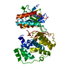
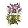
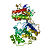
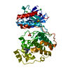
- PDB-4d5a: Clostridial Cysteine protease Cwp84 C116A after propeptide cleavage -
+ Open data

- Basic information
| Entry | Database: PDB / ID: 4d5a | ||||||
|---|---|---|---|---|---|---|---|
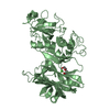
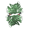
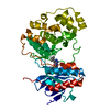
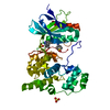
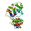
| Title | Clostridial Cysteine protease Cwp84 C116A after propeptide cleavage | ||||||
 Components Components | CELL SURFACE PROTEIN (PUTATIVE CELL SURFACE-ASSOCIATED CYSTEINE PROTEASE) Cell membrane Cell membrane | ||||||
 Keywords Keywords | HYDROLASE / S-LAYER / SURFACE LAYER | ||||||
| Function / homology |  Function and homology information Function and homology information | ||||||
| Biological species |  PEPTOCLOSTRIDIUM DIFFICILE (bacteria) PEPTOCLOSTRIDIUM DIFFICILE (bacteria) | ||||||
| Method | X-RAY DIFFRACTION / SYNCHROTRON / MOLECULAR REPLACEMENT / Resolution: 1.6 Å | ||||||
 Authors Authors | Bradshaw, W.J. / Roberts, A.K. / Shone, C.C. / Acharya, K.R. | ||||||
 Citation Citation | Journal: Acta Crystallogr.,Sect.F / Year: 2015 Title: Cwp84, a Clostridium Difficile Cysteine Protease, Exhibits Conformational Flexibility in the Absence of its Propeptide Authors: Bradshaw, W.J. / Roberts, A.K. / Shone, C.C. / Acharya, K.R. | ||||||
| History |
|
- Structure visualization
| Structure viewer | Molecule: MolmilJmol/JSmol |
|---|
- Downloads & links
-Download
| PDBx/mmCIF format | 4d5a.cif.gz | 196.4 KB | Display | PDBx/mmCIF format |
|---|---|---|---|---|
| PDB format | pdb4d5a.ent.gz | 155.4 KB | Display | PDB format |
| PDBx/mmJSON format | 4d5a.json.gz | Tree view | PDBx/mmJSON format | |
| Others |  Other downloads Other downloads |
-Validation report
| Arichive directory | https://data.pdbj.org/pub/pdb/validation_reports/d5/4d5aftp://data.pdbj.org/pub/pdb/validation_reports/d5/4d5a | HTTPS FTP |
|---|
-Related structure data
| Related structure data |  4d59C 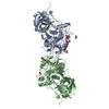 4ci7S C: citing same article ( S: Starting model for refinement |
|---|---|
| Similar structure data |

-Links
 PDBj
PDBj
- Assembly
| Deposited unit | 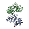
| ||||||||
|---|---|---|---|---|---|---|---|---|---|
| 1 | 
| ||||||||
| 2 | 
| ||||||||
| Unit cell |
|
-Components
| #1: Protein | Cell membrane / CWP84 Mass: 45098.867 Da / Num. of mol.: 2 Fragment: CYSTEINE PROTEASE DOMAIN, LECTIN-LIKE DOMAIN, UNP RESIDUES 92-497 Mutation: YES Source method: isolated from a genetically manipulated source Source: (gene. exp.) PEPTOCLOSTRIDIUM DIFFICILE (bacteria) / Strain: QCD-32G58 / Plasmid: PGEX-6P-1 / Production host: ESCHERICHIA COLI (E. coli) / Strain (production host): BL21(DE3) / References: UniProt: C9YQ11, cathepsin L#2: Chemical | ChemComp-CA /   Mass: 40.078 Da / Num. of mol.: 4 / Source method: obtained synthetically / Formula: Ca Mass: 40.078 Da / Num. of mol.: 4 / Source method: obtained synthetically / Formula: Ca#3: Chemical | ChemComp-PGE / Polyethylene glycol  Mass: 150.173 Da / Num. of mol.: 8 / Source method: obtained synthetically / Formula: C6H14O4 Mass: 150.173 Da / Num. of mol.: 8 / Source method: obtained synthetically / Formula: C6H14O4#4: Chemical | ChemComp-GOL / Glycerol  Mass: 92.094 Da / Num. of mol.: 4 / Source method: obtained synthetically / Formula: C3H8O3 Mass: 92.094 Da / Num. of mol.: 4 / Source method: obtained synthetically / Formula: C3H8O3#5: Water | ChemComp-HOH / | Water Mass: 18.015 Da / Num. of mol.: 791 / Source method: isolated from a natural source / Formula: H2O Mass: 18.015 Da / Num. of mol.: 791 / Source method: isolated from a natural source / Formula: H2OSequence details | STRUCTURE CONSISTS OF RESIDUES 92-497 WITH C116A. THE SIGNAL PEPTIDE (1-32) AND CELL WALL BINDING ...STRUCTURE CONSISTS OF RESIDUES 92-497 WITH C116A. THE SIGNAL PEPTIDE (1-32) AND CELL WALL BINDING DOMAINS (498- 803) WERE NOT PRESENT IN THE CONSTRUCT, THE PROPEPTIDE | |
|---|
-Experimental details
-Experiment
| Experiment | Method: X-RAY DIFFRACTION / Number of used crystals: 1 |
|---|
- Sample preparation
| Crystal | Density Matthews: 2.63 Å3/Da / Density % sol: 53.32 % / Description: NONE |
|---|---|
| Crystal grow | pH: 8 Details: 0.18 M NACL, 90 MM TRIS PH 8.0, 18% PEG 6000, 0.02% W/V 1,4-DIAMINOBUTANE, 0.02% W/V CYSTAMINE DIHYDROCHLORIDE, 0.02% W/V DILOXANIDE FUROATE, 0.02% W/V SARCOSINE, 0.02% W/V SPERMINE, 2MM SODIUM HEPES PH 6.8 |
-Data collection
| Diffraction | Mean temperature: 100 K |
|---|---|
| Diffraction source | Source: SYNCHROTRON / Site: Diamond  / Beamline: I04 / Wavelength: 0.9795 / Beamline: I04 / Wavelength: 0.9795 |
| Detector | Type: DECTRIS PILATUS 6M / Detector: PIXEL / Date: Jan 31, 2014 |
| Radiation | Monochromator: DOUBLE SILICON CRYSTALS / Protocol: SINGLE WAVELENGTH / Monochromatic (M) / Laue (L): M / Scattering type: x-ray |
| Radiation wavelength | Wavelength: 0.9795 Å / Relative weight: 1 |
| Reflection | Resolution: 1.6→47.3 Å / Num. obs: 110175 / % possible obs: 90.8 % / Observed criterion σ(I): 1 / Redundancy: 2.8 % / Rmerge(I) obs: 0.09 / Net I/σ(I): 6.8 |
| Reflection shell | Resolution: 1.6→1.63 Å / Redundancy: 2.5 % / Rmerge(I) obs: 0.47 / Mean I/σ(I) obs: 2.1 / % possible all: 49.9 |
- Processing
| Software |
| ||||||||||||||||||||||||||||||||||||||||||||||||||||||||||||||||||||||||||||||||||||||||||||||||||||||||||||||||||||||||||||||||||||||||||||||||||||||||||||||||||||||||||||||||||||||
|---|---|---|---|---|---|---|---|---|---|---|---|---|---|---|---|---|---|---|---|---|---|---|---|---|---|---|---|---|---|---|---|---|---|---|---|---|---|---|---|---|---|---|---|---|---|---|---|---|---|---|---|---|---|---|---|---|---|---|---|---|---|---|---|---|---|---|---|---|---|---|---|---|---|---|---|---|---|---|---|---|---|---|---|---|---|---|---|---|---|---|---|---|---|---|---|---|---|---|---|---|---|---|---|---|---|---|---|---|---|---|---|---|---|---|---|---|---|---|---|---|---|---|---|---|---|---|---|---|---|---|---|---|---|---|---|---|---|---|---|---|---|---|---|---|---|---|---|---|---|---|---|---|---|---|---|---|---|---|---|---|---|---|---|---|---|---|---|---|---|---|---|---|---|---|---|---|---|---|---|---|---|---|---|
| Refinement | Method to determine structure: MOLECULAR REPLACEMENT Starting model: PDB ENTRY 4CI7 Resolution: 1.6→71.38 Å / Cor.coef. Fo:Fc: 0.949 / Cor.coef. Fo:Fc free: 0.934 / SU B: 2.026 / SU ML: 0.07 / Cross valid method: THROUGHOUT / ESU R: 0.096 / ESU R Free: 0.095 / Stereochemistry target values: MAXIMUM LIKELIHOOD Details: HYDROGENS HAVE BEEN ADDED IN THE RIDING POSITIONS. U VALUES REFINED INDIVIDUALLY
| ||||||||||||||||||||||||||||||||||||||||||||||||||||||||||||||||||||||||||||||||||||||||||||||||||||||||||||||||||||||||||||||||||||||||||||||||||||||||||||||||||||||||||||||||||||||
| Solvent computation | Ion probe radii: 0.8 Å / Shrinkage radii: 0.8 Å / VDW probe radii: 1.2 Å / Solvent model: MASK | ||||||||||||||||||||||||||||||||||||||||||||||||||||||||||||||||||||||||||||||||||||||||||||||||||||||||||||||||||||||||||||||||||||||||||||||||||||||||||||||||||||||||||||||||||||||
| Displacement parameters | Biso mean: 15.346 Å2
| ||||||||||||||||||||||||||||||||||||||||||||||||||||||||||||||||||||||||||||||||||||||||||||||||||||||||||||||||||||||||||||||||||||||||||||||||||||||||||||||||||||||||||||||||||||||
| Refinement step | Cycle: LAST / Resolution: 1.6→71.38 Å
| ||||||||||||||||||||||||||||||||||||||||||||||||||||||||||||||||||||||||||||||||||||||||||||||||||||||||||||||||||||||||||||||||||||||||||||||||||||||||||||||||||||||||||||||||||||||
| Refine LS restraints |
|