 Movie
Movie Controller

[English] 日本語
 Yorodumi
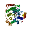
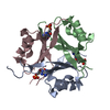
- PDB-4d1f: Crystal structure of the fiber head domain of the Atadenovirus sn... -
+ Open data

- Basic information
| Entry | Database: PDB / ID: 4d1f | ||||||
|---|---|---|---|---|---|---|---|
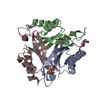
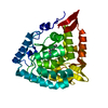
| Title | Crystal structure of the fiber head domain of the Atadenovirus snake adenovirus 1, native, first P212121 crystal form | ||||||
 Components Components | FIBER PROTEIN Fibrous protein Fibrous protein | ||||||
 Keywords Keywords | VIRAL PROTEIN | ||||||
| Function / homology | Adenovirus fibre protein / Attachment protein shaft domain superfamily / viral capsid / cell adhesion / symbiont entry into host cell / host cell nucleus / virion attachment to host cell / Fiber protein Function and homology information Function and homology information | ||||||
| Biological species |  SNAKE ADENOVIRUS 1 SNAKE ADENOVIRUS 1 | ||||||
| Method | X-RAY DIFFRACTION / SYNCHROTRON / MOLECULAR REPLACEMENT / Resolution: 2.7 Å | ||||||
 Authors Authors | Singh, A.K. / van Raaij, M.J. | ||||||
 Citation Citation | Journal: PLoS ONE / Year: 2014 Title: Crystal structure of the fibre head domain of the Atadenovirus Snake Adenovirus 1. Authors: Singh, A.K. / Menendez-Conejero, R. / San Martin, C. / van Raaij, M.J. #1: Journal: Acta Crystallogr. Sect. F Struct. Biol. Cryst. Commun. Year: 2013 Title: Crystallization of the C-terminal domain of the fibre protein from snake adenovirus 1, an atadenovirus. Authors: Singh, A.K. / Menendez-Conejero, R. / San Martin, C. / van Raaij, M.J. #2: Journal: Archives of Virology / Year: 1993 Title: Physicochemical Properties and Cytopathogenicity of an Adenovirus-Like Agent Isolated from Corn Snake (Elaphe Guttata). Authors: Juhasz, A. / Ahne, W. #3: Journal: Virus Res. / Year: 2008 Title: Completion of the Genome Analysis of Snake Adenovirus Type 1, a Representative of the Reptilian Lineage within the Novel Genus Atadenovirus. Authors: Farkas, S.L. / Harrach, B. / Benko, M. | ||||||
| History |
|
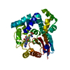
- Structure visualization
| Structure viewer | Molecule: MolmilJmol/JSmol |
|---|
- Downloads & links
-Download
| PDBx/mmCIF format | 4d1f.cif.gz | 252.6 KB | Display | PDBx/mmCIF format |
|---|---|---|---|---|
| PDB format | pdb4d1f.ent.gz | 201.6 KB | Display | PDB format |
| PDBx/mmJSON format | 4d1f.json.gz | Tree view | PDBx/mmJSON format | |
| Others |  Other downloads Other downloads |
-Validation report
| Arichive directory | https://data.pdbj.org/pub/pdb/validation_reports/d1/4d1fftp://data.pdbj.org/pub/pdb/validation_reports/d1/4d1f | HTTPS FTP |
|---|
-Related structure data
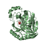
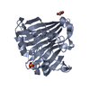
| Related structure data |  4d0uC 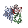 4d0vSC 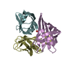 4d1gC 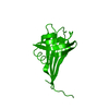 4umiC  4d1h C: citing same article ( S: Starting model for refinement |
|---|---|
| Similar structure data |


-Links
 PDBj
PDBj
- Assembly
| Deposited unit | 
| |||||||||||||||||||||||||||||||||||||||||||||||||||||||||||||||||||||||||||||||||||||||||||||||||||||||||||||||||||||||||||||||||||||||||||||||||||||||||||||||||||||||||||||||||
|---|---|---|---|---|---|---|---|---|---|---|---|---|---|---|---|---|---|---|---|---|---|---|---|---|---|---|---|---|---|---|---|---|---|---|---|---|---|---|---|---|---|---|---|---|---|---|---|---|---|---|---|---|---|---|---|---|---|---|---|---|---|---|---|---|---|---|---|---|---|---|---|---|---|---|---|---|---|---|---|---|---|---|---|---|---|---|---|---|---|---|---|---|---|---|---|---|---|---|---|---|---|---|---|---|---|---|---|---|---|---|---|---|---|---|---|---|---|---|---|---|---|---|---|---|---|---|---|---|---|---|---|---|---|---|---|---|---|---|---|---|---|---|---|---|---|---|---|---|---|---|---|---|---|---|---|---|---|---|---|---|---|---|---|---|---|---|---|---|---|---|---|---|---|---|---|---|---|---|
| 1 | 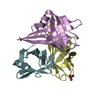
| |||||||||||||||||||||||||||||||||||||||||||||||||||||||||||||||||||||||||||||||||||||||||||||||||||||||||||||||||||||||||||||||||||||||||||||||||||||||||||||||||||||||||||||||||
| 2 | 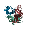
| |||||||||||||||||||||||||||||||||||||||||||||||||||||||||||||||||||||||||||||||||||||||||||||||||||||||||||||||||||||||||||||||||||||||||||||||||||||||||||||||||||||||||||||||||
| 3 | 
| |||||||||||||||||||||||||||||||||||||||||||||||||||||||||||||||||||||||||||||||||||||||||||||||||||||||||||||||||||||||||||||||||||||||||||||||||||||||||||||||||||||||||||||||||
| 4 | 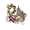
| |||||||||||||||||||||||||||||||||||||||||||||||||||||||||||||||||||||||||||||||||||||||||||||||||||||||||||||||||||||||||||||||||||||||||||||||||||||||||||||||||||||||||||||||||
| Unit cell |
| |||||||||||||||||||||||||||||||||||||||||||||||||||||||||||||||||||||||||||||||||||||||||||||||||||||||||||||||||||||||||||||||||||||||||||||||||||||||||||||||||||||||||||||||||
| Noncrystallographic symmetry (NCS) | NCS domain:
NCS domain segments: Ens-ID: 1 / Refine code: 1
|