 Movie
Movie Controller

[English] 日本語
 Yorodumi
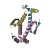
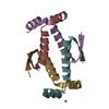
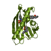
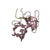
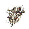
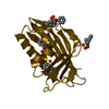
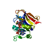
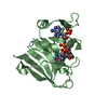
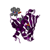
- PDB-4cz7: Truncated tetramerization domain of zebrafish p53 (crystal form III) -
+ Open data

- Basic information
| Entry | Database: PDB / ID: 4cz7 | ||||||
|---|---|---|---|---|---|---|---|
| Title | Truncated tetramerization domain of zebrafish p53 (crystal form III) | ||||||
 Components Components | CELLULAR TUMOR ANTIGEN P53 P53 P53 | ||||||
 Keywords Keywords | CELL CYCLE / TUMOR SUPPRESSOR / TRANSCRIPTION FACTOR PROTEIN EVOLUTION / DANIO RERIO | ||||||
| Function / homology |  Function and homology informationregulation of oogenesis / Autodegradation of the E3 ubiquitin ligase COP1 / Regulation of TP53 Expression / Regulation of TP53 Activity through Acetylation / Regulation of TP53 Activity through Association with Co-factors / Regulation of TP53 Activity through Methylation / G2/M Checkpoints / RUNX3 regulates CDKN1A transcription / PKR-mediated signaling / Ovarian tumor domain proteases ...regulation of oogenesis / Autodegradation of the E3 ubiquitin ligase COP1 / Regulation of TP53 Expression / Regulation of TP53 Activity through Acetylation / Regulation of TP53 Activity through Association with Co-factors / Regulation of TP53 Activity through Methylation / G2/M Checkpoints / RUNX3 regulates CDKN1A transcription / PKR-mediated signaling / Ovarian tumor domain proteases / Oxidative Stress Induced Senescence / Oncogene Induced Senescence / Regulation of TP53 Activity through Phosphorylation / Regulation of TP53 Degradation / Stabilization of p53 / epithelium development / The role of GTSE1 in G2/M progression after G2 checkpoint / Ub-specific processing proteases / programmed cell death involved in cell development / intestinal epithelial structure maintenance / hematopoietic stem cell homeostasis / epidermis morphogenesis / apoptotic process involved in blood vessel morphogenesis / negative regulation of cell division / retrotransposon silencing / regulation of double-strand break repair / apoptotic process involved in development / response to methylmercury / definitive hemopoiesis / regulation of intrinsic apoptotic signaling pathway by p53 class mediator / DNA damage response, signal transduction by p53 class mediator resulting in transcription of p21 class mediator / hematopoietic stem cell proliferation / erythrocyte maturation / erythrocyte development / negative regulation of cell cycle / response to X-ray / intrinsic apoptotic signaling pathway in response to DNA damage by p53 class mediator / anatomical structure morphogenesis / positive regulation of cell cycle / response to UV / negative regulation of angiogenesis / intrinsic apoptotic signaling pathway / transforming growth factor beta receptor signaling pathway / DNA damage checkpoint signaling / promoter-specific chromatin binding / protein tetramerization / regulation of protein stability / autophagy / cellular senescence / positive regulation of neuron apoptotic process / regulation of cell population proliferation / cellular response to hypoxia / spermatogenesis / sequence-specific DNA binding / transcription cis-regulatory region binding / DNA-binding transcription factor activity, RNA polymerase II-specific / positive regulation of apoptotic process / cell cycle / RNA polymerase II cis-regulatory region sequence-specific DNA binding / DNA-binding transcription factor activity / apoptotic process / DNA damage response / chromatin / regulation of transcription by RNA polymerase II / positive regulation of DNA-templated transcription / positive regulation of transcription by RNA polymerase II / mitochondrion / identical protein binding / metal ion binding / nucleus / cytoplasm Function and homology informationregulation of oogenesis / Autodegradation of the E3 ubiquitin ligase COP1 / Regulation of TP53 Expression / Regulation of TP53 Activity through Acetylation / Regulation of TP53 Activity through Association with Co-factors / Regulation of TP53 Activity through Methylation / G2/M Checkpoints / RUNX3 regulates CDKN1A transcription / PKR-mediated signaling / Ovarian tumor domain proteases ...regulation of oogenesis / Autodegradation of the E3 ubiquitin ligase COP1 / Regulation of TP53 Expression / Regulation of TP53 Activity through Acetylation / Regulation of TP53 Activity through Association with Co-factors / Regulation of TP53 Activity through Methylation / G2/M Checkpoints / RUNX3 regulates CDKN1A transcription / PKR-mediated signaling / Ovarian tumor domain proteases / Oxidative Stress Induced Senescence / Oncogene Induced Senescence / Regulation of TP53 Activity through Phosphorylation / Regulation of TP53 Degradation / Stabilization of p53 / epithelium development / The role of GTSE1 in G2/M progression after G2 checkpoint / Ub-specific processing proteases / programmed cell death involved in cell development / intestinal epithelial structure maintenance / hematopoietic stem cell homeostasis / epidermis morphogenesis / apoptotic process involved in blood vessel morphogenesis / negative regulation of cell division / retrotransposon silencing / regulation of double-strand break repair / apoptotic process involved in development / response to methylmercury / definitive hemopoiesis / regulation of intrinsic apoptotic signaling pathway by p53 class mediator / DNA damage response, signal transduction by p53 class mediator resulting in transcription of p21 class mediator / hematopoietic stem cell proliferation / erythrocyte maturation / erythrocyte development / negative regulation of cell cycle / response to X-ray / intrinsic apoptotic signaling pathway in response to DNA damage by p53 class mediator / anatomical structure morphogenesis / positive regulation of cell cycle / response to UV / negative regulation of angiogenesis / intrinsic apoptotic signaling pathway / transforming growth factor beta receptor signaling pathway / DNA damage checkpoint signaling / promoter-specific chromatin binding / protein tetramerization / regulation of protein stability / autophagy / cellular senescence / positive regulation of neuron apoptotic process / regulation of cell population proliferation / cellular response to hypoxia / spermatogenesis / sequence-specific DNA binding / transcription cis-regulatory region binding / DNA-binding transcription factor activity, RNA polymerase II-specific / positive regulation of apoptotic process / cell cycle / RNA polymerase II cis-regulatory region sequence-specific DNA binding / DNA-binding transcription factor activity / apoptotic process / DNA damage response / chromatin / regulation of transcription by RNA polymerase II / positive regulation of DNA-templated transcription / positive regulation of transcription by RNA polymerase II / mitochondrion / identical protein binding / metal ion binding / nucleus / cytoplasmSimilarity search - Function | ||||||
| Biological species |  DANIO RERIO (zebrafish) DANIO RERIO (zebrafish) | ||||||
| Method | X-RAY DIFFRACTION / SYNCHROTRON / MOLECULAR REPLACEMENT / Resolution: 1.1 Å | ||||||
 Authors Authors | Joerger, A.C. | ||||||
 Citation Citation | Journal: Structure / Year: 2014 Title: Tracing the Evolution of the P53 Tetramerization Domain Authors: Joerger, A.C. / Wilcken, R. / Andreeva, A. | ||||||
| History |
|
- Structure visualization
| Structure viewer | Molecule: MolmilJmol/JSmol |
|---|
- Downloads & links
-Download
| PDBx/mmCIF format | 4cz7.cif.gz | 93.5 KB | Display | PDBx/mmCIF format |
|---|---|---|---|---|
| PDB format | pdb4cz7.ent.gz | 75.3 KB | Display | PDB format |
| PDBx/mmJSON format | 4cz7.json.gz | Tree view | PDBx/mmJSON format | |
| Others |  Other downloads Other downloads |
-Validation report
| Arichive directory | https://data.pdbj.org/pub/pdb/validation_reports/cz/4cz7ftp://data.pdbj.org/pub/pdb/validation_reports/cz/4cz7 | HTTPS FTP |
|---|
-Related structure data


-Links
 PDBj
PDBj


- Assembly
| Deposited unit | 
| ||||||||
|---|---|---|---|---|---|---|---|---|---|
| 1 | 
| ||||||||
| 2 | 
| ||||||||
| Unit cell |
|
-Components
| #1: Protein/peptide | P53 Mass: 3799.270 Da / Num. of mol.: 6 Fragment: TRUNCATED TETRAMERIZATION DOMAIN, RESIDUES 302-331 Source method: isolated from a genetically manipulated source Source: (gene. exp.) DANIO RERIO (zebrafish) / Production host:  ESCHERICHIA COLI (E. coli) / References: UniProt: G1K2L5, UniProt: P79734*PLUS ESCHERICHIA COLI (E. coli) / References: UniProt: G1K2L5, UniProt: P79734*PLUS#2: Chemical | Phosphate  Mass: 94.971 Da / Num. of mol.: 3 / Source method: obtained synthetically / Formula: PO4 Mass: 94.971 Da / Num. of mol.: 3 / Source method: obtained synthetically / Formula: PO4#3: Chemical | ChemComp-GOL / | Glycerol  Mass: 92.094 Da / Num. of mol.: 1 / Source method: obtained synthetically / Formula: C3H8O3 Mass: 92.094 Da / Num. of mol.: 1 / Source method: obtained synthetically / Formula: C3H8O3#4: Water | ChemComp-HOH / | Water Mass: 18.015 Da / Num. of mol.: 140 / Source method: isolated from a natural source / Formula: H2O Mass: 18.015 Da / Num. of mol.: 140 / Source method: isolated from a natural source / Formula: H2OSequence details | ADDITIONAL | |
|---|
-Experimental details
-Experiment
| Experiment | Method: X-RAY DIFFRACTION / Number of used crystals: 1 |
|---|
- Sample preparation
| Crystal | Density Matthews: 2 Å3/Da / Density % sol: 38 % / Description: NONE |
|---|---|
| Crystal grow | Temperature: 293 K / Method: vapor diffusion, sitting drop / pH: 5 Details: SITTING DROP VAPOR DIFFUSION AT 20 DEGREE C; PROTEIN SOLUTION: 17 MG/ML IN 20 MM TRIS PH 7.5, 50 MM NACL, 5 MM DTT; CRYSTALLIZATION BUFFER: 1.8 M SODIUM/POTASSIUM PHOSPHATE, PH 5.0. |
-Data collection
| Diffraction | Mean temperature: 100 K |
|---|---|
| Diffraction source | Source: SYNCHROTRON / Site: Diamond  / Beamline: I03 / Wavelength: 0.8266 / Beamline: I03 / Wavelength: 0.8266 |
| Radiation | Protocol: SINGLE WAVELENGTH / Monochromatic (M) / Laue (L): M / Scattering type: x-ray |
| Radiation wavelength | Wavelength: 0.8266 Å / Relative weight: 1 |
| Reflection | Resolution: 1.1→27.7 Å / Num. obs: 68831 / % possible obs: 94.8 % / Observed criterion σ(I): 2 / Redundancy: 3.5 % / Rmerge(I) obs: 0.06 / Net I/σ(I): 12.1 |
| Reflection shell | Resolution: 1.1→1.16 Å / Redundancy: 3.3 % / Rmerge(I) obs: 0.17 / Mean I/σ(I) obs: 6.5 / % possible all: 87.4 |
- Processing
| Software | Name: REFMAC / Version: 5.8.0069 / Classification: refinement | ||||||||||||||||||||||||||||||||||||||||||||||||||||||||||||||||||||||||||||||||||||||||||||||||||||||||||||||||||||||||||||||||||||||||||||||||||||||||||||||||||||||||||||||||||||||
|---|---|---|---|---|---|---|---|---|---|---|---|---|---|---|---|---|---|---|---|---|---|---|---|---|---|---|---|---|---|---|---|---|---|---|---|---|---|---|---|---|---|---|---|---|---|---|---|---|---|---|---|---|---|---|---|---|---|---|---|---|---|---|---|---|---|---|---|---|---|---|---|---|---|---|---|---|---|---|---|---|---|---|---|---|---|---|---|---|---|---|---|---|---|---|---|---|---|---|---|---|---|---|---|---|---|---|---|---|---|---|---|---|---|---|---|---|---|---|---|---|---|---|---|---|---|---|---|---|---|---|---|---|---|---|---|---|---|---|---|---|---|---|---|---|---|---|---|---|---|---|---|---|---|---|---|---|---|---|---|---|---|---|---|---|---|---|---|---|---|---|---|---|---|---|---|---|---|---|---|---|---|---|---|
| Refinement | Method to determine structure: MOLECULAR REPLACEMENT / Resolution: 1.1→27.7 Å / Cor.coef. Fo:Fc: 0.972 / Cor.coef. Fo:Fc free: 0.967 / SU B: 0.789 / SU ML: 0.018 / Cross valid method: THROUGHOUT / ESU R: 0.031 / ESU R Free: 0.031 / Stereochemistry target values: MAXIMUM LIKELIHOOD / Details: HYDROGENS HAVE BEEN ADDED IN THE RIDING POSITIONS.
| ||||||||||||||||||||||||||||||||||||||||||||||||||||||||||||||||||||||||||||||||||||||||||||||||||||||||||||||||||||||||||||||||||||||||||||||||||||||||||||||||||||||||||||||||||||||
| Solvent computation | Ion probe radii: 0.8 Å / Shrinkage radii: 0.8 Å / VDW probe radii: 1.2 Å / Solvent model: MASK | ||||||||||||||||||||||||||||||||||||||||||||||||||||||||||||||||||||||||||||||||||||||||||||||||||||||||||||||||||||||||||||||||||||||||||||||||||||||||||||||||||||||||||||||||||||||
| Displacement parameters | Biso mean: 12.823 Å2
| ||||||||||||||||||||||||||||||||||||||||||||||||||||||||||||||||||||||||||||||||||||||||||||||||||||||||||||||||||||||||||||||||||||||||||||||||||||||||||||||||||||||||||||||||||||||
| Refinement step | Cycle: LAST / Resolution: 1.1→27.7 Å
| ||||||||||||||||||||||||||||||||||||||||||||||||||||||||||||||||||||||||||||||||||||||||||||||||||||||||||||||||||||||||||||||||||||||||||||||||||||||||||||||||||||||||||||||||||||||
| Refine LS restraints |
|