 Movie
Movie Controller

[English] 日本語
 Yorodumi
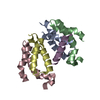
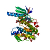
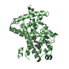
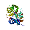
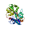
- PDB-4cyc: CRYSTAL STRUCTURE OF A UBX-EXD-DNA COMPLEX INCLUDING THE HEXAPEPT... -
+ Open data

- Basic information
| Entry | Database: PDB / ID: 4cyc | ||||||
|---|---|---|---|---|---|---|---|
| Title | CRYSTAL STRUCTURE OF A UBX-EXD-DNA COMPLEX INCLUDING THE HEXAPEPTIDE AND UBDA MOTIFS | ||||||
 Components Components |
| ||||||
 Keywords Keywords |  TRANSCRIPTION / HOX / PBC / DNA PROTEIN COMPLEX TRANSCRIPTION / HOX / PBC / DNA PROTEIN COMPLEX | ||||||
| Function / homology |  Function and homology information Function and homology informationdorsal vessel aortic cell fate commitment / positive regulation of muscle organ development / anterior Malpighian tubule development / specification of animal organ identity / regulation of imaginal disc growth / salivary gland boundary specification / haltere development / oenocyte development / mesodermal cell fate specification / specification of segmental identity, thorax ...dorsal vessel aortic cell fate commitment / positive regulation of muscle organ development / anterior Malpighian tubule development / specification of animal organ identity / regulation of imaginal disc growth / salivary gland boundary specification / haltere development / oenocyte development / mesodermal cell fate specification / specification of segmental identity, thorax / open tracheal system development / imaginal disc-derived leg morphogenesis / somatic muscle development / muscle cell fate specification / polytene chromosome band / endoderm formation / eye development / regulation of cell fate specification / peripheral nervous system development / cell fate determination / anterior/posterior pattern specification / midgut development / transcription factor binding / neuron development / embryonic organ development / cis-regulatory region sequence-specific DNA binding / transcription repressor complex / transcription coregulator binding / protein-DNA complex / transcription coregulator activity / animal organ morphogenesis / brain development / RNA polymerase II transcription regulator complex / heart development / DNA-binding transcription factor binding / transcription regulator complex / sequence-specific DNA binding / transcription coactivator activity / DNA-binding transcription factor activity, RNA polymerase II-specific / RNA polymerase II cis-regulatory region sequence-specific DNA binding / DNA-binding transcription factor activity / protein heterodimerization activity / protein domain specific binding / negative regulation of DNA-templated transcription / regulation of DNA-templated transcription / regulation of transcription by RNA polymerase II / positive regulation of DNA-templated transcription / negative regulation of transcription by RNA polymerase II / positive regulation of transcription by RNA polymerase II / DNA binding / nucleus / cytoplasmSimilarity search - Function | ||||||
| Biological species |  DROSOPHILA MELANOGASTER (fruit fly) DROSOPHILA MELANOGASTER (fruit fly) | ||||||
| Method | X-RAY DIFFRACTION / SYNCHROTRON / MOLECULAR REPLACEMENT / Resolution: 2.36 Å | ||||||
 Authors Authors | Foos, N. / Mate, M.J. / Ortiz-Lombardia, M. | ||||||
 Citation Citation | Journal: Structure / Year: 2015 Title: A Flexible Extension of the Drosophila Ultrabithorax Homeodomain Defines a Novel Hox/Pbc Interaction Mode. Authors: Foos, N. / Maurel-Zaffran, C. / Mate, M.J. / Vincentelli, R. / Hainaut, M. / Berenger, H. / Pradel, J. / Saurin, A.J. / Ortiz-Lombardia, M. / Graba, Y. | ||||||
| History |
|
- Structure visualization
| Structure viewer | Molecule: MolmilJmol/JSmol |
|---|
- Downloads & links
-Download
| PDBx/mmCIF format | 4cyc.cif.gz | 106.9 KB | Display | PDBx/mmCIF format |
|---|---|---|---|---|
| PDB format | pdb4cyc.ent.gz | 80.3 KB | Display | PDB format |
| PDBx/mmJSON format | 4cyc.json.gz | Tree view | PDBx/mmJSON format | |
| Others |  Other downloads Other downloads |
-Validation report
| Arichive directory | https://data.pdbj.org/pub/pdb/validation_reports/cy/4cycftp://data.pdbj.org/pub/pdb/validation_reports/cy/4cyc | HTTPS FTP |
|---|
-Related structure data
| Related structure data | 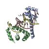 4uusC  4uutC  4bzl S: Starting model for refinement C: citing same article ( |
|---|---|
| Similar structure data |


-Links
 PDBj
PDBj


- Assembly
| Deposited unit | 
| ||||||||
|---|---|---|---|---|---|---|---|---|---|
| 1 |
| ||||||||
| Unit cell |
|
-Components
| #1: Protein | Homeosis Mass: 11459.246 Da / Num. of mol.: 1 / Fragment: HOMEODOMAIN WITH HX AND UBDA, RESIDUES 233-367 Source method: isolated from a genetically manipulated source Source: (gene. exp.) DROSOPHILA MELANOGASTER (fruit fly) / Plasmid: UBXIVA PDEST14 / Production host:  ESCHERICHIA COLI (E. coli) / Strain (production host): BL21(DE3) / Variant (production host): ROSETTA PLYSS / References: UniProt: P83949 ESCHERICHIA COLI (E. coli) / Strain (production host): BL21(DE3) / Variant (production host): ROSETTA PLYSS / References: UniProt: P83949 |
|---|---|
| #2: Protein | / DPBX / HOMEOTIC PROTEIN EXTRADENTICLE Mass: 8857.946 Da / Num. of mol.: 1 / Fragment: HOMEODOMAIN RESIDUES 238-312 Source method: isolated from a genetically manipulated source Source: (gene. exp.) DROSOPHILA MELANOGASTER (fruit fly) / Plasmid: EXD2 PETG20A / Production host: ESCHERICHIA COLI (E. coli) / Strain (production host): BL21(DE3) / Variant (production host): ROSETTA PLYSS / References: UniProt: P40427 |
| #3: DNA chain | Mass: 4537.975 Da / Num. of mol.: 1 / Source method: obtained synthetically / Source: (synth.) DROSOPHILA MELANOGASTER (fruit fly) |
| #4: DNA chain | Mass: 4640.020 Da / Num. of mol.: 1 / Source method: obtained synthetically / Source: (synth.) DROSOPHILA MELANOGASTER (fruit fly) |
| #5: Water | ChemComp-HOH / Water Mass: 18.015 Da / Num. of mol.: 68 / Source method: isolated from a natural source / Formula: H2O Mass: 18.015 Da / Num. of mol.: 68 / Source method: isolated from a natural source / Formula: H2O |
| Sequence details | FIRST METHIONINE |
-Experimental details
-Experiment
| Experiment | Method: X-RAY DIFFRACTION / Number of used crystals: 1 |
|---|
- Sample preparation
| Crystal | Density Matthews: 3.48 Å3/Da / Density % sol: 68.5 % / Description: NONE |
|---|---|
| Crystal grow | pH: 7.5 / Details: 0.1 M AMMONIUM PHOSPHATE, pH 7.5 |
-Data collection
| Diffraction | Mean temperature: 100 K |
|---|---|
| Diffraction source | Source: SYNCHROTRON / Site: SOLEIL  / Beamline: PROXIMA 1 / Wavelength: 1.3853 / Beamline: PROXIMA 1 / Wavelength: 1.3853 |
| Detector | Type: DECTRIS PILATUS 6M / Detector: PIXEL / Date: Feb 3, 2012 |
| Radiation | Protocol: SINGLE WAVELENGTH / Monochromatic (M) / Laue (L): M / Scattering type: x-ray |
| Radiation wavelength | Wavelength: 1.3853 Å / Relative weight: 1 |
| Reflection | Resolution: 2.36→84.62 Å / Num. obs: 17094 / % possible obs: 100 % / Observed criterion σ(I): -3.7 / Redundancy: 10.8 % / Biso Wilson estimate: 83.97 Å2 / Rmerge(I) obs: 0.04 / Net I/σ(I): 41.2 |
| Reflection shell | Resolution: 2.36→2.37 Å / Redundancy: 10.8 % / Rmerge(I) obs: 0.97 / Mean I/σ(I) obs: 2.4 / % possible all: 100 |
- Processing
| Software |
| |||||||||||||||||||||||||||||||||||||||||||||||||||||||||||||||||||||||||||||||||||||||||||||||||||||||||||||||||||||||||||||
|---|---|---|---|---|---|---|---|---|---|---|---|---|---|---|---|---|---|---|---|---|---|---|---|---|---|---|---|---|---|---|---|---|---|---|---|---|---|---|---|---|---|---|---|---|---|---|---|---|---|---|---|---|---|---|---|---|---|---|---|---|---|---|---|---|---|---|---|---|---|---|---|---|---|---|---|---|---|---|---|---|---|---|---|---|---|---|---|---|---|---|---|---|---|---|---|---|---|---|---|---|---|---|---|---|---|---|---|---|---|---|---|---|---|---|---|---|---|---|---|---|---|---|---|---|---|---|
| Refinement | Method to determine structure: MOLECULAR REPLACEMENT Starting model: PDB ENTRY 4BZL 4bzl Resolution: 2.36→25 Å / Cor.coef. Fo:Fc: 0.9425 / Cor.coef. Fo:Fc free: 0.9429 / SU R Cruickshank DPI: 0.21 / Cross valid method: THROUGHOUT / σ(F): 0 / SU R Blow DPI: 0.218 / SU Rfree Blow DPI: 0.174 / SU Rfree Cruickshank DPI: 0.171
| |||||||||||||||||||||||||||||||||||||||||||||||||||||||||||||||||||||||||||||||||||||||||||||||||||||||||||||||||||||||||||||
| Displacement parameters | Biso mean: 87.18 Å2
| |||||||||||||||||||||||||||||||||||||||||||||||||||||||||||||||||||||||||||||||||||||||||||||||||||||||||||||||||||||||||||||
| Refine analyze | Luzzati coordinate error obs: 0.828 Å | |||||||||||||||||||||||||||||||||||||||||||||||||||||||||||||||||||||||||||||||||||||||||||||||||||||||||||||||||||||||||||||
| Refinement step | Cycle: LAST / Resolution: 2.36→25 Å
| |||||||||||||||||||||||||||||||||||||||||||||||||||||||||||||||||||||||||||||||||||||||||||||||||||||||||||||||||||||||||||||
| Refine LS restraints |
| |||||||||||||||||||||||||||||||||||||||||||||||||||||||||||||||||||||||||||||||||||||||||||||||||||||||||||||||||||||||||||||
| LS refinement shell | Resolution: 2.36→2.5 Å / Total num. of bins used: 9
| |||||||||||||||||||||||||||||||||||||||||||||||||||||||||||||||||||||||||||||||||||||||||||||||||||||||||||||||||||||||||||||
| Refinement TLS params. | Method: refined / Refine-ID: X-RAY DIFFRACTION
| |||||||||||||||||||||||||||||||||||||||||||||||||||||||||||||||||||||||||||||||||||||||||||||||||||||||||||||||||||||||||||||
| Refinement TLS group |
|
