 Movie
Movie Controller

[English] 日本語
 Yorodumi
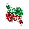
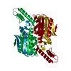
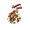
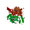
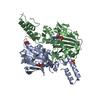
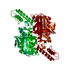
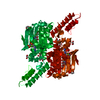
- PDB-4cs2: Catalytic domain of Pyrrolysyl-tRNA synthetase mutant Y306A, Y384... -
+ Open data

- Basic information
| Entry | Database: PDB / ID: 4cs2 | ||||||
|---|---|---|---|---|---|---|---|
| Title | Catalytic domain of Pyrrolysyl-tRNA synthetase mutant Y306A, Y384F in its apo form | ||||||
 Components Components | PYRROLYSINE--TRNA LIGASE | ||||||
 Keywords Keywords |  LIGASE LIGASE | ||||||
| Function / homology |  Function and homology information Function and homology informationpyrrolysine-tRNAPyl ligase / pyrrolysyl-tRNA synthetase activity / tRNA aminoacylation for protein translation / tRNA binding / ATP binding / cytoplasmSimilarity search - Function | ||||||
| Biological species |  METHANOSARCINA MAZEI (archaea) METHANOSARCINA MAZEI (archaea) | ||||||
| Method | X-RAY DIFFRACTION / SYNCHROTRON / MOLECULAR REPLACEMENT / Resolution: 1.9 Å | ||||||
 Authors Authors | Schmidt, M.J. / Weber, A. / Pott, M. / Welte, W. / Summerer, D. | ||||||
 Citation Citation | Journal: Chembiochem / Year: 2014 Title: Structural Basis of Furan-Amino Acid Recognition by a Polyspecific Aminoacyl-tRNA-Synthetase and its Genetic Encoding in Human Cells. Authors: Schmidt, M.J. / Weber, A. / Pott, M. / Welte, W. / Summerer, D. | ||||||
| History |
|
- Structure visualization
| Structure viewer | Molecule: MolmilJmol/JSmol |
|---|
- Downloads & links
-Download
| PDBx/mmCIF format | 4cs2.cif.gz | 120.9 KB | Display | PDBx/mmCIF format |
|---|---|---|---|---|
| PDB format | pdb4cs2.ent.gz | 93 KB | Display | PDB format |
| PDBx/mmJSON format | 4cs2.json.gz | Tree view | PDBx/mmJSON format | |
| Others |  Other downloads Other downloads |
-Validation report
| Arichive directory | https://data.pdbj.org/pub/pdb/validation_reports/cs/4cs2ftp://data.pdbj.org/pub/pdb/validation_reports/cs/4cs2 | HTTPS FTP |
|---|
-Related structure data
| Related structure data |  4cs3C 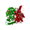 4cs4C 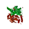 2q7eS S: Starting model for refinement C: citing same article ( |
|---|---|
| Similar structure data |


-Links
 PDBj
PDBj
- Assembly
| Deposited unit | 
| ||||||||
|---|---|---|---|---|---|---|---|---|---|
| 1 | 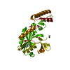
| ||||||||
| Unit cell |
|
-Components
| #1: Protein | Mass: 31736.404 Da / Num. of mol.: 1 / Fragment: C-TERMINAL FRAGMENT, RESIDUES 188-454 / Mutation: YES Source method: isolated from a genetically manipulated source Source: (gene. exp.) METHANOSARCINA MAZEI (archaea) / Production host:  ESCHERICHIA COLI (E. coli) / Variant (production host): TOP10 / References: UniProt: Q8PWY1, pyrrolysine-tRNAPyl ligase ESCHERICHIA COLI (E. coli) / Variant (production host): TOP10 / References: UniProt: Q8PWY1, pyrrolysine-tRNAPyl ligase | ||
|---|---|---|---|
| #2: Chemical | ChemComp-PEG / Diethylene glycol  Mass: 106.120 Da / Num. of mol.: 1 / Source method: obtained synthetically / Formula: C4H10O3 Mass: 106.120 Da / Num. of mol.: 1 / Source method: obtained synthetically / Formula: C4H10O3 | ||
| #3: Chemical | ChemComp-EDO / Ethylene glycol  Mass: 62.068 Da / Num. of mol.: 4 / Source method: obtained synthetically / Formula: C2H6O2 Mass: 62.068 Da / Num. of mol.: 4 / Source method: obtained synthetically / Formula: C2H6O2#4: Water | ChemComp-HOH / | Water Mass: 18.015 Da / Num. of mol.: 126 / Source method: isolated from a natural source / Formula: H2O Mass: 18.015 Da / Num. of mol.: 126 / Source method: isolated from a natural source / Formula: H2O |
-Experimental details
-Experiment
| Experiment | Method: X-RAY DIFFRACTION / Number of used crystals: 1 |
|---|
- Sample preparation
| Crystal | Density Matthews: 2.23 Å3/Da / Density % sol: 44.96 % / Description: NONE |
|---|---|
| Crystal grow | pH: 8.5 Details: 50MM TRIS-HCL PH 8.5, 200 MM CALCIUM CHLORIDE AND 18 % (W/V) PEG4000 |
-Data collection
| Diffraction | Mean temperature: 100 K |
|---|---|
| Diffraction source | Source: SYNCHROTRON / Site: SLS  / Beamline: X06SA / Wavelength: 0.9999 / Beamline: X06SA / Wavelength: 0.9999 |
| Detector | Type: DECTRIS PILATUS 6M / Detector: PIXEL / Date: Feb 23, 2013 |
| Radiation | Protocol: SINGLE WAVELENGTH / Monochromatic (M) / Laue (L): M / Scattering type: x-ray |
| Radiation wavelength | Wavelength: 0.9999 Å / Relative weight: 1 |
| Reflection | Resolution: 1.9→39.92 Å / Num. obs: 21692 / % possible obs: 99.3 % / Redundancy: 3.3 % / Biso Wilson estimate: 31.67 Å2 / Rmerge(I) obs: 0.04 / Net I/σ(I): 18.75 |
| Reflection shell | Resolution: 1.9→1.97 Å / Redundancy: 3.2 % / Rmerge(I) obs: 0.49 / Mean I/σ(I) obs: 2.5 / % possible all: 98.5 |
- Processing
| Software |
| ||||||||||||||||||||||||||||||||||||||||||||||||||||||||||||||||||||||||||||||||||||||||||||||||||||
|---|---|---|---|---|---|---|---|---|---|---|---|---|---|---|---|---|---|---|---|---|---|---|---|---|---|---|---|---|---|---|---|---|---|---|---|---|---|---|---|---|---|---|---|---|---|---|---|---|---|---|---|---|---|---|---|---|---|---|---|---|---|---|---|---|---|---|---|---|---|---|---|---|---|---|---|---|---|---|---|---|---|---|---|---|---|---|---|---|---|---|---|---|---|---|---|---|---|---|---|---|---|
| Refinement | Method to determine structure: MOLECULAR REPLACEMENT Starting model: PDB ENTRY 2Q7E Resolution: 1.9→39.924 Å / SU ML: 0.21 / σ(F): 1.36 / Phase error: 25.26 / Stereochemistry target values: ML
| ||||||||||||||||||||||||||||||||||||||||||||||||||||||||||||||||||||||||||||||||||||||||||||||||||||
| Solvent computation | Shrinkage radii: 0.9 Å / VDW probe radii: 1.11 Å / Solvent model: FLAT BULK SOLVENT MODEL | ||||||||||||||||||||||||||||||||||||||||||||||||||||||||||||||||||||||||||||||||||||||||||||||||||||
| Displacement parameters | Biso mean: 43.1 Å2 | ||||||||||||||||||||||||||||||||||||||||||||||||||||||||||||||||||||||||||||||||||||||||||||||||||||
| Refinement step | Cycle: LAST / Resolution: 1.9→39.924 Å
| ||||||||||||||||||||||||||||||||||||||||||||||||||||||||||||||||||||||||||||||||||||||||||||||||||||
| Refine LS restraints |
| ||||||||||||||||||||||||||||||||||||||||||||||||||||||||||||||||||||||||||||||||||||||||||||||||||||
| LS refinement shell |
| ||||||||||||||||||||||||||||||||||||||||||||||||||||||||||||||||||||||||||||||||||||||||||||||||||||
| Refinement TLS params. | Method: refined / Refine-ID: X-RAY DIFFRACTION
| ||||||||||||||||||||||||||||||||||||||||||||||||||||||||||||||||||||||||||||||||||||||||||||||||||||
| Refinement TLS group |
|
