 Movie
Movie Controller

[English] 日本語
 Yorodumi
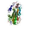
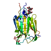
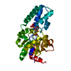
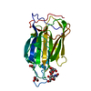
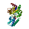
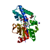
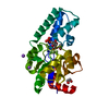
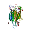
- PDB-4cou: Crystal Structure of Epithelial Adhesin 6 A domain (Epa6A) from C... -
+ Open data

- Basic information
| Entry | Database: PDB / ID: 4cou | |||||||||
|---|---|---|---|---|---|---|---|---|---|---|
| Title | Crystal Structure of Epithelial Adhesin 6 A domain (Epa6A) from Candida glabrata in complex with Lactose | |||||||||
 Components Components | EPITHELIAL ADHESIN 6 | |||||||||
 Keywords Keywords |  CELL ADHESION / LECTIN / TISSUE INVASION / PATHOGENICITY CELL ADHESION / LECTIN / TISSUE INVASION / PATHOGENICITY | |||||||||
| Function / homology |  Function and homology information Function and homology informationcell-abiotic substrate adhesion / single-species biofilm formation on inanimate substrate / fungal biofilm matrix / adhesion of symbiont to host / fungal-type cell wall / carbohydrate binding / cell surface / extracellular region / metal ion bindingSimilarity search - Function | |||||||||
| Biological species |  CANDIDA GLABRATA (fungus) CANDIDA GLABRATA (fungus) | |||||||||
| Method | X-RAY DIFFRACTION / SYNCHROTRON / MOLECULAR REPLACEMENT / Resolution: 1.48 Å | |||||||||
 Authors Authors | Kock, M. / Maestre-Reyna, M. / Diderrich, R. / Moesch, H.-U. / Essen, L.-O. | |||||||||
 Citation Citation | Journal: J.Biol.Chem. / Year: 2015 Title: Structural Hotspots Determine Functional Diversity of the Candida Glabrata Epithelial Adhesin Family Authors: Diderrich, R. / Kock, M. / Maestre-Reyna, M. / Rupp, S. / Essen, L.-O. / Moesch, H.-U. | |||||||||
| History |
|
- Structure visualization
| Structure viewer | Molecule: MolmilJmol/JSmol |
|---|
- Downloads & links
-Download
| PDBx/mmCIF format | 4cou.cif.gz | 120.7 KB | Display | PDBx/mmCIF format |
|---|---|---|---|---|
| PDB format | pdb4cou.ent.gz | 91.3 KB | Display | PDB format |
| PDBx/mmJSON format | 4cou.json.gz | Tree view | PDBx/mmJSON format | |
| Others |  Other downloads Other downloads |
-Validation report
| Arichive directory | https://data.pdbj.org/pub/pdb/validation_reports/co/4couftp://data.pdbj.org/pub/pdb/validation_reports/co/4cou | HTTPS FTP |
|---|
-Related structure data
| Related structure data | 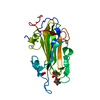 4covC  4cowC 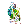 4coyC  4cozC 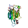 4d3wC 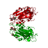 4af9S C: citing same article ( S: Starting model for refinement |
|---|---|
| Similar structure data |


-Links
 PDBj
PDBj


- Assembly
| Deposited unit | 
| ||||||||
|---|---|---|---|---|---|---|---|---|---|
| 1 |
| ||||||||
| Unit cell |
|
-Components
-Protein / Sugars , 2 types, 2 molecules A
| #1: Protein | Mass: 29937.945 Da / Num. of mol.: 1 / Fragment: ADHESION DOMAIN (A DOMAIN), RESIDUES 26-271 Source method: isolated from a genetically manipulated source Source: (gene. exp.) CANDIDA GLABRATA (fungus) / Strain: CBS138 / Plasmid: PET28A / Production host:  ESCHERICHIA COLI B (bacteria) / Variant (production host): SHUFFLE T7 EXPRESS (C3029) / References: UniProt: Q6FX55 ESCHERICHIA COLI B (bacteria) / Variant (production host): SHUFFLE T7 EXPRESS (C3029) / References: UniProt: Q6FX55 |
|---|---|
| #2: Polysaccharide | beta-D-galactopyranose-(1-4)-beta-D-glucopyranose / beta-lactose  , Oligosaccharide / Class: Nutrient / Mass: 342.297 Da / Num. of mol.: 1 , Oligosaccharide / Class: Nutrient / Mass: 342.297 Da / Num. of mol.: 1Source method: isolated from a genetically manipulated source Details: oligosaccharide / References: beta-lactose |
-Non-polymers , 4 types, 248 molecules 


| #3: Chemical | ChemComp-CA /  Mass: 40.078 Da / Num. of mol.: 1 / Source method: obtained synthetically / Formula: Ca Mass: 40.078 Da / Num. of mol.: 1 / Source method: obtained synthetically / Formula: Ca |
|---|---|
| #4: Chemical | ChemComp-ACT / Acetate Mass: 59.044 Da / Num. of mol.: 1 / Source method: obtained synthetically / Formula: C2H3O2 Mass: 59.044 Da / Num. of mol.: 1 / Source method: obtained synthetically / Formula: C2H3O2 |
| #5: Chemical | ChemComp-GOL / Glycerol Mass: 92.094 Da / Num. of mol.: 1 / Source method: obtained synthetically / Formula: C3H8O3 Mass: 92.094 Da / Num. of mol.: 1 / Source method: obtained synthetically / Formula: C3H8O3 |
| #6: Water | ChemComp-HOH / WaterMass: 18.015 Da / Num. of mol.: 245 / Source method: isolated from a natural source / Formula: H2O |
-Experimental details
-Experiment
| Experiment | Method: X-RAY DIFFRACTION / Number of used crystals: 1 |
|---|
- Sample preparation
| Crystal | Density Matthews: 2.24 Å3/Da / Density % sol: 45.14 % / Description: NONE |
|---|---|
| Crystal grow | Temperature: 291 K / Method: vapor diffusion, sitting drop Details: 0.08 M SODIUM ACETATE PH:4.6, 1.6 M AMMONIUM SULFATE, 20% GLYCEROL, 0.05 M LACTOSE, 291 K, VAPOR DIFFUSION IN SITTING DROP |
-Data collection
| Diffraction | Mean temperature: 100 K |
|---|---|
| Diffraction source | Source: SYNCHROTRON / Site: BESSY  / Beamline: 14.1 / Wavelength: 0.91841 / Beamline: 14.1 / Wavelength: 0.91841 |
| Detector | Type: MARRESEARCH MARMOSAIC 255 MM / Detector: CCD / Date: Sep 16, 2012 |
| Radiation | Monochromator: KMC-1 / Protocol: SINGLE WAVELENGTH / Monochromatic (M) / Laue (L): M / Scattering type: x-ray |
| Radiation wavelength | Wavelength: 0.91841 Å / Relative weight: 1 |
| Reflection | Resolution: 1.48→28.77 Å / Num. obs: 45573 / % possible obs: 99.7 % / Observed criterion σ(I): -3 / Redundancy: 3.8 % / Rmerge(I) obs: 0.04 / Net I/σ(I): 21.1 |
| Reflection shell | Resolution: 1.48→1.56 Å / Redundancy: 3.8 % / Rmerge(I) obs: 0.57 / Mean I/σ(I) obs: 2.9 / % possible all: 99.9 |
- Processing
| Software |
| ||||||||||||||||||||||||||||||||||||||||||||||||||||||||||||||||||||||||||||||||||||||||||||||||||||||||||||||||||||||||||||||||||||||||||||||||||||||||||||||||||||||||||||||||||||||
|---|---|---|---|---|---|---|---|---|---|---|---|---|---|---|---|---|---|---|---|---|---|---|---|---|---|---|---|---|---|---|---|---|---|---|---|---|---|---|---|---|---|---|---|---|---|---|---|---|---|---|---|---|---|---|---|---|---|---|---|---|---|---|---|---|---|---|---|---|---|---|---|---|---|---|---|---|---|---|---|---|---|---|---|---|---|---|---|---|---|---|---|---|---|---|---|---|---|---|---|---|---|---|---|---|---|---|---|---|---|---|---|---|---|---|---|---|---|---|---|---|---|---|---|---|---|---|---|---|---|---|---|---|---|---|---|---|---|---|---|---|---|---|---|---|---|---|---|---|---|---|---|---|---|---|---|---|---|---|---|---|---|---|---|---|---|---|---|---|---|---|---|---|---|---|---|---|---|---|---|---|---|---|---|
| Refinement | Method to determine structure: MOLECULAR REPLACEMENT Starting model: PRUNED VERSION OF PDB ENTRY 4AF9 Resolution: 1.48→26.5 Å / Cor.coef. Fo:Fc: 0.974 / Cor.coef. Fo:Fc free: 0.97 / SU B: 2.189 / SU ML: 0.037 / Cross valid method: THROUGHOUT / ESU R: 0.062 / ESU R Free: 0.056 / Stereochemistry target values: MAXIMUM LIKELIHOOD Details: HYDROGENS HAVE BEEN ADDED IN THE RIDING POSITIONS. U VALUES REFINED INDIVIDUALLY
| ||||||||||||||||||||||||||||||||||||||||||||||||||||||||||||||||||||||||||||||||||||||||||||||||||||||||||||||||||||||||||||||||||||||||||||||||||||||||||||||||||||||||||||||||||||||
| Solvent computation | Ion probe radii: 0.8 Å / Shrinkage radii: 0.8 Å / VDW probe radii: 1.2 Å / Solvent model: MASK | ||||||||||||||||||||||||||||||||||||||||||||||||||||||||||||||||||||||||||||||||||||||||||||||||||||||||||||||||||||||||||||||||||||||||||||||||||||||||||||||||||||||||||||||||||||||
| Displacement parameters | Biso mean: 19.167 Å2
| ||||||||||||||||||||||||||||||||||||||||||||||||||||||||||||||||||||||||||||||||||||||||||||||||||||||||||||||||||||||||||||||||||||||||||||||||||||||||||||||||||||||||||||||||||||||
| Refinement step | Cycle: LAST / Resolution: 1.48→26.5 Å
| ||||||||||||||||||||||||||||||||||||||||||||||||||||||||||||||||||||||||||||||||||||||||||||||||||||||||||||||||||||||||||||||||||||||||||||||||||||||||||||||||||||||||||||||||||||||
| Refine LS restraints |
|