reduction of food intake in response to dietary excess / RecQ family helicase-topoisomerase III complex / DNA topoisomerase activity / resolution of DNA recombination intermediates / chromosome separation / DNA topoisomerase / DNA topoisomerase type I (single strand cut, ATP-independent) activity / resolution of meiotic recombination intermediates / mitochondrial DNA metabolic process / Impaired BRCA2 binding to PALB2 ...reduction of food intake in response to dietary excess / RecQ family helicase-topoisomerase III complex / DNA topoisomerase activity / resolution of DNA recombination intermediates / chromosome separation / DNA topoisomerase / DNA topoisomerase type I (single strand cut, ATP-independent) activity / resolution of meiotic recombination intermediates / mitochondrial DNA metabolic process / Impaired BRCA2 binding to PALB2 / Defective homologous recombination repair (HRR) due to BRCA1 loss of function / Defective HDR through Homologous Recombination Repair (HRR) due to PALB2 loss of BRCA1 binding function / Defective HDR through Homologous Recombination Repair (HRR) due to PALB2 loss of BRCA2/RAD51/RAD51C binding function / Homologous DNA Pairing and Strand Exchange / Resolution of D-loop Structures through Synthesis-Dependent Strand Annealing (SDSA) / Resolution of D-loop Structures through Holliday Junction Intermediates / HDR through Single Strand Annealing (SSA) / Impaired BRCA2 binding to RAD51 / DNA topological change / Presynaptic phase of homologous DNA pairing and strand exchange / response to glucose / meiotic cell cycle / double-strand break repair via homologous recombination / HDR through Homologous Recombination (HRR) / G2/M DNA damage checkpoint / multicellular organism growth / PML body / Meiotic recombination / glucose homeostasis / single-stranded DNA binding / Processing of DNA double-strand break ends / Regulation of TP53 Activity through Phosphorylation / DNA replication / nuclear body / mitochondrial matrix / nucleotide binding / DNA binding / zinc ion binding / nucleoplasm / nucleus Similarity search - Function
RecQ-mediated genome instability protein 1, N-terminal domain / RecQ-mediated genome instability protein Rmi1, C-terminal domain / RecQ-mediated genome instability protein 1, C-terminal OB-fold domain / RecQ-mediated genome instability protein 1, N-terminal helical domain superfamily / : / Recq-mediated genome instability protein 1, C-terminal OB-fold / RMI1, N-terminal helical domain / DUF1767 / Zinc finger GRF-type profile. / DNA topoisomerase 3-like, TOPRIM domain ...RecQ-mediated genome instability protein 1, N-terminal domain / RecQ-mediated genome instability protein Rmi1, C-terminal domain / RecQ-mediated genome instability protein 1, C-terminal OB-fold domain / RecQ-mediated genome instability protein 1, N-terminal helical domain superfamily / : / Recq-mediated genome instability protein 1, C-terminal OB-fold / RMI1, N-terminal helical domain / DUF1767 / Zinc finger GRF-type profile. / DNA topoisomerase 3-like, TOPRIM domain / Zinc finger, GRF-type / GRF zinc finger / RecQ mediated genome instability protein 1, N-terminal / RecQ mediated genome instability protein, N-terminal OB-fold superfamily / RMI1, N-terminal OB-fold domain / DNA topoisomerase, type IA, zn finger / Topoisomerase DNA binding C4 zinc finger / Topoisomerase I, domain 3 / Topoisomerase I; domain 2 / Topoisomerase I, domain 2 / Rossmann fold - #140 / Topoisomerase I; domain 4 / Topoisomerase I, domain 4 / DNA topoisomerase, type IA / DNA topoisomerase, type IA, central region, subdomain 2 / DNA topoisomerase, type IA, active site / Topoisomerase (Topo) IA-type active site signature. / Topoisomerase (Topo) IA-type catalytic domain profile. / DNA topoisomerase, type IA, domain 2 / DNA topoisomerase, type IA, DNA-binding domain / DNA topoisomerase, type IA, central / DNA topoisomerase, type IA, central region, subdomain 1 / DNA topoisomerase, type IA, central region, subdomain 3 / DNA topoisomerase, type IA, core domain / DNA topoisomerase / Bacterial DNA topoisomeraes I ATP-binding domain / Bacterial DNA topoisomerase I DNA-binding domain / Topoisomerase I; domain 3 / TOPRIM / Toprim domain / Toprim domain profile. / TOPRIM domain / Helicase, Ruva Protein; domain 3 / Distorted Sandwich / OB fold (Dihydrolipoamide Acetyltransferase, E2P) / Beta Barrel / Rossmann fold / Orthogonal Bundle / 3-Layer(aba) Sandwich / Mainly Beta / Mainly Alpha / Alpha Beta Similarity search - Domain/homology
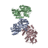
DNATOPOISOMERASE3-ALPHA / DNA TOPOISOMERASE III ALPHA
Mass: 86641.688 Da / Num. of mol.: 1 Source method: isolated from a genetically manipulated source Source: (gene. exp.) HOMO SAPIENS (human) / Cell line (production host): High Five / Production host: TRICHOPLUSIA NI (cabbage looper) / References: UniProt: Q13472, DNA topoisomerase
#2: Protein
RECQ-MEDIATEDGENOMEINSTABILITYPROTEIN1 / BLM-ASSOCIATED PROTEIN OF 75 KDA / BLAP75 / FAAP75
Mass: 24644.457 Da / Num. of mol.: 1 / Fragment: RESIDUES 1-219 Source method: isolated from a genetically manipulated source Source: (gene. exp.) HOMO SAPIENS (human) / Cell line (production host): High Five / Production host: TRICHOPLUSIA NI (cabbage looper) / References: UniProt: Q9H9A7
In the structure databanks used in Yorodumi, some data are registered as the other names, "COVID-19 virus" and "2019-nCoV". Here are the details of the virus and the list of structure data.
Jan 31, 2019. EMDB accession codes are about to change! (news from PDBe EMDB page)
EMDB accession codes are about to change! (news from PDBe EMDB page)
The allocation of 4 digits for EMDB accession codes will soon come to an end. Whilst these codes will remain in use, new EMDB accession codes will include an additional digit and will expand incrementally as the available range of codes is exhausted. The current 4-digit format prefixed with “EMD-” (i.e. EMD-XXXX) will advance to a 5-digit format (i.e. EMD-XXXXX), and so on. It is currently estimated that the 4-digit codes will be depleted around Spring 2019, at which point the 5-digit format will come into force.
The EM Navigator/Yorodumi systems omit the EMD- prefix.
Related info.:Q: What is EMD? / ID/Accession-code notation in Yorodumi/EM Navigator
Yorodumi is a browser for structure data from EMDB, PDB, SASBDB, etc.
This page is also the successor to EM Navigator detail page, and also detail information page/front-end page for Omokage search.
The word "yorodu" (or yorozu) is an old Japanese word meaning "ten thousand". "mi" (miru) is to see.
Related info.:EMDB / PDB / SASBDB / Comparison of 3 databanks / Yorodumi Search / Aug 31, 2016. New EM Navigator & Yorodumi / Yorodumi Papers / Jmol/JSmol / Function and homology information / Changes in new EM Navigator and Yorodumi
 Movie
Movie Controller

 Yorodumi
 Open data
 Basic information
 Components
 Keywords
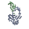
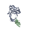
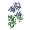
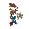
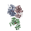
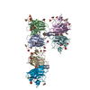
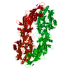
 CELL CYCLE / DOUBLE HOLLIDAY JUNCTION DISSOLUTION / DECATENATION / MINIMAL DISSOLVASOME
 Function and homology information
 Authors
 Citation
 Structure visualization
 Downloads & links
 Other downloads


 PDBj
PDBj


 Assembly
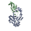


 Mass: 40.078 Da / Num. of mol.: 1 / Source method: obtained synthetically / Formula: Ca
 Mass: 18.015 Da / Num. of mol.: 5 / Source method: isolated from a natural source / Formula: H2O
 Sample preparation
 / Beamline: X10SA / Wavelength: 1
  Processing