E3UBIQUITIN/ISG15LIGASETRIM25 / ESTROGEN-RESPONSIVE FINGER PROTEIN / RING FINGER PROTEIN 147 / TRIPARTITE MOTIF-CONTAINING PROTEIN ...ESTROGEN-RESPONSIVE FINGER PROTEIN / RING FINGER PROTEIN 147 / TRIPARTITE MOTIF-CONTAINING PROTEIN 25 / UBIQUITIN/ISG15-CONJUGATING ENZYME TRIM25 / ZINC FINGER PROTEIN 147 / TRIM
Mass: 71448.484 Da / Num. of mol.: 2 / Mutation: YES Source method: isolated from a genetically manipulated source Source: (gene. exp.) HOMO SAPIENS (human) / Production host: ESCHERICHIA COLI (E. coli) References: UniProt: Q14258, ubiquitin-protein ligase, Ligases; Forming carbon-nitrogen bonds; Acid-amino-acid ligases (peptide synthases)
Method to determine structure: OTHER Starting model: NONE Resolution: 2.8→56.88 Å / Cor.coef. Fo:Fc: 0.937 / Cor.coef. Fo:Fc free: 0.916 / SU B: 48.703 / SU ML: 0.419 / Cross valid method: THROUGHOUT / ESU R Free: 0.5 Stereochemistry target values: MAXIMUM LIKELIHOOD WITH PHASES Details: HYDROGENS HAVE BEEN ADDED IN THE RIDING POSITIONS.
Rfactor
Num. reflection
% reflection
Selection details
Rfree
0.30914
975
10 %
RANDOM
Rwork
0.25106
-
-
-
obs
0.25685
8805
96.06 %
-
Solvent computation
Ion probe radii: 0.8 Å / Shrinkage radii: 0.8 Å / VDW probe radii: 1.2 Å / Solvent model: MASK
 Movie
Movie Controller


 Open data
 Basic information
 Components
 Keywords
 LIGASE
 Function and homology information
 Authors
 Citation
 Structure visualization
 Downloads & links
 Other downloads
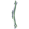


 PDBj
PDBj


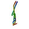
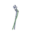
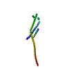
 Assembly

 Mass: 18.015 Da / Num. of mol.: 8 / Source method: isolated from a natural source / Formula: H2O
 Sample preparation
 / Type:
  Processing