 Movie
Movie Controller

[English] 日本語
 Yorodumi
- PDB-4cf7: Crystal structure of adenylate kinase from Aquifex aeolicus with ... -
+ Open data

- Basic information
| Entry | Database: PDB / ID: 4cf7 | ||||||
|---|---|---|---|---|---|---|---|
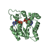
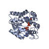
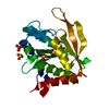
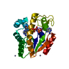
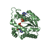
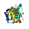
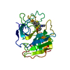
| Title | Crystal structure of adenylate kinase from Aquifex aeolicus with MgADP bound | ||||||
 Components Components | ADENYLATE KINASE | ||||||
 Keywords Keywords | TRANSFERASE / PHOSPHORYL TRANSFER / NUCLEOTIDE-BINDING | ||||||
| Function / homology |  Function and homology information Function and homology informationnucleoside monophosphate metabolic process / nucleoside diphosphate metabolic process / adenylate kinase / adenylate kinase activity / AMP salvage / nucleoside diphosphate kinase activity / phosphorylation / intracellular membrane-bounded organelle / ATP binding / cytosol / cytoplasmSimilarity search - Function | ||||||
| Biological species |   AQUIFEX AEOLICUS (bacteria) AQUIFEX AEOLICUS (bacteria) | ||||||
| Method | X-RAY DIFFRACTION / SYNCHROTRON / MOLECULAR REPLACEMENT / Resolution: 1.594 Å | ||||||
 Authors Authors | Kerns, S.J. / Agafonov, R.V. / Cho, Y.-J. / Pontiggia, F. / Otten, R. / Pachov, D.V. / Kutter, S. / Phung, L.A. / Murphy, P.N. / Thai, V. ...Kerns, S.J. / Agafonov, R.V. / Cho, Y.-J. / Pontiggia, F. / Otten, R. / Pachov, D.V. / Kutter, S. / Phung, L.A. / Murphy, P.N. / Thai, V. / Hagan, M.F. / Kern, D. | ||||||
 Citation Citation | Journal: Nat.Struct.Mol.Biol. / Year: 2015 Title: The Energy Landscape of Adenylate Kinase During Catalysis. Authors: Kerns, S.J. / Agafonov, R.V. / Cho, Y. / Pontiggia, F. / Otten, R. / Pachov, D.V. / Kutter, S. / Phung, L.A. / Murphy, P.N. / Thai, V. / Alber, T. / Hagan, M.F. / Kern, D. | ||||||
| History |
|
- Structure visualization
| Structure viewer | Molecule: MolmilJmol/JSmol |
|---|
- Downloads & links
-Download
| PDBx/mmCIF format | 4cf7.cif.gz | 106.9 KB | Display | PDBx/mmCIF format |
|---|---|---|---|---|
| PDB format | pdb4cf7.ent.gz | 81.5 KB | Display | PDB format |
| PDBx/mmJSON format | 4cf7.json.gz | Tree view | PDBx/mmJSON format | |
| Others |  Other downloads Other downloads |
-Validation report
| Arichive directory | https://data.pdbj.org/pub/pdb/validation_reports/cf/4cf7ftp://data.pdbj.org/pub/pdb/validation_reports/cf/4cf7 | HTTPS FTP |
|---|
-Related structure data
| Related structure data | 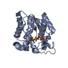 3sr0SC  4jkyC 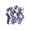 4jl5C  4jl6C  4jl8C  4jlaC  4jlbC 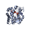 4jldC 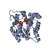 4jloC 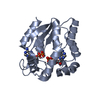 4jlpC S: Starting model for refinement C: citing same article ( |
|---|---|
| Similar structure data |


-Links
 PDBj
PDBj


- Assembly
| Deposited unit | 
| ||||||||
|---|---|---|---|---|---|---|---|---|---|
| 1 | 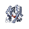
| ||||||||
| 2 | 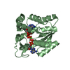
| ||||||||
| Unit cell |
|
-Components
| #1: Protein | / AK / ATP-AMP TRANSPHOSPHORYLASE / ATP\:AMP PHOSPHOTRANSFERASE / ADENYLATE MONOPHOSPHATE KINASE Mass: 23269.057 Da / Num. of mol.: 2 Source method: isolated from a genetically manipulated source Source: (gene. exp.) AQUIFEX AEOLICUS (bacteria) / Production host: ESCHERICHIA COLI (E. coli) / Strain (production host): BL21(DE3) / References: UniProt: O66490, adenylate kinase#2: Chemical | ChemComp-ADP / Adenosine diphosphate  Mass: 427.201 Da / Num. of mol.: 4 / Source method: obtained synthetically / Formula: C10H15N5O10P2 / Comment: ADP, energy-carrying molecule*YM Mass: 427.201 Da / Num. of mol.: 4 / Source method: obtained synthetically / Formula: C10H15N5O10P2 / Comment: ADP, energy-carrying molecule*YM#3: Chemical | ChemComp-AMP / | Adenosine monophosphate  Mass: 347.221 Da / Num. of mol.: 1 / Source method: obtained synthetically / Formula: C10H14N5O7P / Comment: AMP*YM Mass: 347.221 Da / Num. of mol.: 1 / Source method: obtained synthetically / Formula: C10H14N5O7P / Comment: AMP*YM#4: Chemical | ChemComp-MG / |   Mass: 24.305 Da / Num. of mol.: 1 / Source method: obtained synthetically / Formula: Mg Mass: 24.305 Da / Num. of mol.: 1 / Source method: obtained synthetically / Formula: Mg#5: Water | ChemComp-HOH / | Water Mass: 18.015 Da / Num. of mol.: 334 / Source method: isolated from a natural source / Formula: H2O Mass: 18.015 Da / Num. of mol.: 334 / Source method: isolated from a natural source / Formula: H2O |
|---|
-Experimental details
-Experiment
| Experiment | Method: X-RAY DIFFRACTION / Number of used crystals: 1 |
|---|
- Sample preparation
| Crystal | Density Matthews: 1.95 Å3/Da / Density % sol: 37.02 % / Description: NONE |
|---|---|
| Crystal grow | Temperature: 291 K / Method: vapor diffusion, sitting drop / pH: 9 Details: A 1.5 UL SOLUTION CONTAINING 26 MG/ML OF PROTEIN, 20 MM MGCL2 AND 20 MM ADP IN 50 MM TRIS-HCL PH 7 WAS MIXED WITH 0.2 M AMMONIUM ACETATE, 0.1 M SODIUM ACETATE TRIHYDRATE PH 5.6, 30% W/V PEG- ...Details: A 1.5 UL SOLUTION CONTAINING 26 MG/ML OF PROTEIN, 20 MM MGCL2 AND 20 MM ADP IN 50 MM TRIS-HCL PH 7 WAS MIXED WITH 0.2 M AMMONIUM ACETATE, 0.1 M SODIUM ACETATE TRIHYDRATE PH 5.6, 30% W/V PEG-4000 IN A 1:1 RATIO; VAPOR DIFFUSION; SITTING DROP; 291 K |
-Data collection
| Diffraction | Mean temperature: 100 K |
|---|---|
| Diffraction source | Source: SYNCHROTRON / Site: ALS  / Beamline: 8.2.1 / Wavelength: 1.00001 / Beamline: 8.2.1 / Wavelength: 1.00001 |
| Detector | Type: ADSC QUANTUM 315r / Detector: CCD / Date: Jun 12, 2013 |
| Radiation | Monochromator: DOUBLE CRYSTAL . SI(111) / Protocol: SINGLE WAVELENGTH / Monochromatic (M) / Laue (L): M / Scattering type: x-ray |
| Radiation wavelength | Wavelength: 1.00001 Å / Relative weight: 1 |
| Reflection | Resolution: 1.59→64.7 Å / Num. obs: 48649 / % possible obs: 100 % / Observed criterion σ(I): 3.1 / Redundancy: 6.5 % / Biso Wilson estimate: 15.53 Å2 / Rmerge(I) obs: 0.09 / Net I/σ(I): 12.2 |
| Reflection shell | Resolution: 1.59→1.64 Å / Redundancy: 6.7 % / Rmerge(I) obs: 0.62 / Mean I/σ(I) obs: 3.1 / % possible all: 100 |
- Processing
| Software |
| ||||||||||||||||||||||||||||||||||||||||||||||||||||||||
|---|---|---|---|---|---|---|---|---|---|---|---|---|---|---|---|---|---|---|---|---|---|---|---|---|---|---|---|---|---|---|---|---|---|---|---|---|---|---|---|---|---|---|---|---|---|---|---|---|---|---|---|---|---|---|---|---|---|
| Refinement | Method to determine structure: MOLECULAR REPLACEMENT Starting model: PDB ENTRY 3SR0 Resolution: 1.594→40.265 Å / SU ML: 0.15 / σ(F): 1.34 / Phase error: 22.77 / Stereochemistry target values: ML
| ||||||||||||||||||||||||||||||||||||||||||||||||||||||||
| Solvent computation | Shrinkage radii: 0.9 Å / VDW probe radii: 1.11 Å / Solvent model: FLAT BULK SOLVENT MODEL | ||||||||||||||||||||||||||||||||||||||||||||||||||||||||
| Displacement parameters | Biso mean: 20.8 Å2 | ||||||||||||||||||||||||||||||||||||||||||||||||||||||||
| Refinement step | Cycle: LAST / Resolution: 1.594→40.265 Å
| ||||||||||||||||||||||||||||||||||||||||||||||||||||||||
| Refine LS restraints |
| ||||||||||||||||||||||||||||||||||||||||||||||||||||||||
| LS refinement shell |
|
