| Entry | Database: PDB / ID: 4c7v
|
|---|
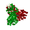
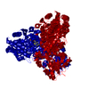
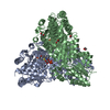
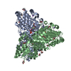
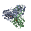
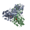
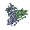
| Title | Apo Transketolase from Lactobacillus salivarius at 2.2A resolution |
|---|
 Components Components | TRANSKETOLASE |
|---|
 Keywords Keywords | TRANSFERASE |
|---|
| Function / homology |  Function and homology information Function and homology information
Transketolase, bacterial-like / Transketolase family / Transketolase signature 1. / Transketolase, thiamine diphosphate binding domain / Transketolase, N-terminal / Transketolase, C-terminal domain / Transketolase, C-terminal domain / Rossmann fold - #920 / Transketolase-like, pyrimidine-binding domain / Transketolase, pyrimidine binding domain ...Transketolase, bacterial-like / Transketolase family / Transketolase signature 1. / Transketolase, thiamine diphosphate binding domain / Transketolase, N-terminal / Transketolase, C-terminal domain / Transketolase, C-terminal domain / Rossmann fold - #920 / Transketolase-like, pyrimidine-binding domain / Transketolase, pyrimidine binding domain / Transketolase, pyrimidine binding domain / Transketolase C-terminal/Pyruvate-ferredoxin oxidoreductase domain II / Thiamin diphosphate (ThDP)-binding fold, Pyr/PP domains / Thiamin diphosphate-binding fold / Rossmann fold / 3-Layer(aba) Sandwich / Alpha BetaSimilarity search - Domain/homology |
|---|
| Biological species |  LACTOBACILLUS SALIVARIUS (bacteria) LACTOBACILLUS SALIVARIUS (bacteria) |
|---|
| Method | X-RAY DIFFRACTION / SYNCHROTRON / MOLECULAR REPLACEMENT / Resolution: 2.2 Å |
|---|
 Authors Authors | Lobley, C.M.C. / Lukacik, P. / Bumann, M. / Aller, P. / Douangamath, A. / O'Toole, P.W. / Walsh, M.A. |
|---|
 Citation Citation | Journal: Acta Crystallogr.,Sect.F / Year: 2015
Title: High Resolution Crystal Structures of Lactobacillus Salivarius Transketolase in the Presence and Absence of Thiamine Pyrophosphate
Authors: Lukacik, P. / Lobley, C.M.C. / Bumann, M. / Arena De Souza, V. / Owens, R.J. / O'Toole, P.W. / Walsh, M.A. |
|---|
| History | | Deposition | Sep 26, 2013 | Deposition site: PDBE / Processing site: PDBE |
|---|
| Revision 1.0 | Oct 8, 2014 | Provider: repository / Type: Initial release |
|---|
| Revision 1.1 | Oct 14, 2015 | Group: Database references / Structure summary |
|---|
| Revision 1.2 | Oct 21, 2015 | Group: Database references |
|---|
| Revision 1.3 | May 8, 2024 | Group: Data collection / Database references / Other
Category: chem_comp_atom / chem_comp_bond ...chem_comp_atom / chem_comp_bond / database_2 / pdbx_database_status
Item: _database_2.pdbx_DOI / _database_2.pdbx_database_accession / _pdbx_database_status.status_code_sf |
|---|
|
|---|
 Movie
Movie Controller

 Yorodumi
 Open data
 Basic information
 Structure visualization
 Downloads & links
 Other downloads


 PDBj
PDBj


 Assembly
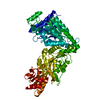

 Mass: 18.015 Da / Num. of mol.: 246 / Source method: isolated from a natural source / Formula: H2O
 Sample preparation
 / Beamline: I04-1 / Wavelength: 0.917
  Processing