 Movie
Movie Controller


+ Open data

- Basic information
| Entry | Database: PDB / ID: 4c0t | ||||||
|---|---|---|---|---|---|---|---|
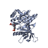
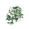
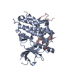
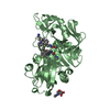
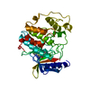
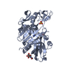
| Title | Candida albicans PKh Kinase Domain | ||||||
 Components Components | LIKELY PROTEIN KINASE | ||||||
 Keywords Keywords |  TRANSFERASE / PDK1 ORTHOLOG TRANSFERASE / PDK1 ORTHOLOG | ||||||
| Function / homology |  Function and homology informationendocytosis / cell cortex / peptidyl-serine phosphorylation / non-specific serine/threonine protein kinase / intracellular signal transduction / protein serine/threonine kinase activity / lipid binding / ATP binding / nucleus Function and homology informationendocytosis / cell cortex / peptidyl-serine phosphorylation / non-specific serine/threonine protein kinase / intracellular signal transduction / protein serine/threonine kinase activity / lipid binding / ATP binding / nucleusSimilarity search - Function | ||||||
| Biological species |  CANDIDA ALBICANS (yeast) CANDIDA ALBICANS (yeast) | ||||||
| Method | X-RAY DIFFRACTION / SYNCHROTRON / MOLECULAR REPLACEMENT / Resolution: 3.16 Å | ||||||
 Authors Authors | Schulze, J.O. / Pastor-Flores, D. / Biondi, R.M. | ||||||
 Citation Citation | Journal: Acs Chem.Biol. / Year: 2013 Title: The Pif-Pocket as a Target for C. Albicans Pkh Selective Inhibitors. Authors: Pastor-Flores, D. / Schulze, J.O. / Bahi, A. / Giacometti, R. / Ferrer-Dalmau, J. / Passeron, S. / Engel, M. / Suess, E. / Casamayor, A. / Biondi, R.M. | ||||||
| History |
| ||||||
| Remark 650 | HELIX DETERMINATION METHOD: AUTHOR PROVIDED. | ||||||
| Remark 700 | SHEET DETERMINATION METHOD: AUTHOR PROVIDED. |
- Structure visualization
| Structure viewer | Molecule: MolmilJmol/JSmol |
|---|
- Downloads & links
-Download
| PDBx/mmCIF format | 4c0t.cif.gz | 85.9 KB | Display | PDBx/mmCIF format |
|---|---|---|---|---|
| PDB format | pdb4c0t.ent.gz | 56.8 KB | Display | PDB format |
| PDBx/mmJSON format | 4c0t.json.gz | Tree view | PDBx/mmJSON format | |
| Others |  Other downloads Other downloads |
-Validation report
| Arichive directory | https://data.pdbj.org/pub/pdb/validation_reports/c0/4c0tftp://data.pdbj.org/pub/pdb/validation_reports/c0/4c0t | HTTPS FTP |
|---|
-Related structure data
| Related structure data |  3hrcS S: Starting model for refinement |
|---|---|
| Similar structure data |


-Links
 PDBj
PDBj- Assembly
| Deposited unit | 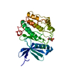
| ||||||||
|---|---|---|---|---|---|---|---|---|---|
| 1 |
| ||||||||
| Unit cell |
|
-Components
| #1: Protein | Mass: 110088.344 Da / Num. of mol.: 1 Source method: isolated from a genetically manipulated source Source: (gene. exp.) CANDIDA ALBICANS (yeast) / Strain: SC5314Description: SYNTHETIC GENE BECAUSE CANDIDA HAS NON-UNIVERSAL CODON USAGE Plasmid: PFASTBAC / Production host:   SPODOPTERA FRUGIPERDA (fall armyworm) SPODOPTERA FRUGIPERDA (fall armyworm)References: UniProt: Q5A3P6, non-specific serine/threonine protein kinase |
|---|
-Experimental details
-Experiment
| Experiment | Method: X-RAY DIFFRACTION / Number of used crystals: 1 |
|---|
- Sample preparation
| Crystal | Density Matthews: 2.2 Å3/Da / Density % sol: 45 % / Description: NONE |
|---|---|
| Crystal grow | pH: 5.6 Details: 100 MM AMMONIUM SULFATE, 30 % (W/V) PEG 4000, AND 100 MM SODIUM CITRATE/ CITRIC ACID (PH 5.6) |
-Data collection
| Diffraction | Mean temperature: 100 K |
|---|---|
| Diffraction source | Source: SYNCHROTRON / Site: BESSY  / Beamline: 14.1 / Wavelength: 0.91841 / Beamline: 14.1 / Wavelength: 0.91841 |
| Detector | Type: MARRESEARCH / Detector: CCD / Date: Jul 30, 2011 / Details: MIRRORS |
| Radiation | Monochromator: SI-111 CRYSTAL / Protocol: SINGLE WAVELENGTH / Monochromatic (M) / Laue (L): M / Scattering type: x-ray |
| Radiation wavelength | Wavelength: 0.91841 Å / Relative weight: 1 |
| Reflection | Resolution: 3.16→70.9 Å / Num. obs: 5590 / % possible obs: 100 % / Observed criterion σ(I): 3 / Redundancy: 7.8 % / Biso Wilson estimate: 53.51 Å2 / Rmerge(I) obs: 0.17 / Net I/σ(I): 14.54 |
| Reflection shell | Resolution: 3.16→3.26 Å / Redundancy: 8 % / Rmerge(I) obs: 0.8 / Mean I/σ(I) obs: 3 / % possible all: 100 |
- Processing
| Software |
| ||||||||||||||||||||||||
|---|---|---|---|---|---|---|---|---|---|---|---|---|---|---|---|---|---|---|---|---|---|---|---|---|---|
| Refinement | Method to determine structure: MOLECULAR REPLACEMENT Starting model: PDB ENTRY 3HRC Resolution: 3.16→70.859 Å / SU ML: 0.45 / σ(F): 2.02 / Phase error: 27.09 / Stereochemistry target values: ML
| ||||||||||||||||||||||||
| Solvent computation | Shrinkage radii: 0.8 Å / VDW probe radii: 1 Å / Solvent model: FLAT BULK SOLVENT MODEL | ||||||||||||||||||||||||
| Displacement parameters | Biso mean: 32.1 Å2 | ||||||||||||||||||||||||
| Refinement step | Cycle: LAST / Resolution: 3.16→70.859 Å
| ||||||||||||||||||||||||
| Refine LS restraints |
| ||||||||||||||||||||||||
| LS refinement shell |
|
