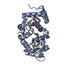
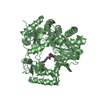
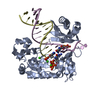
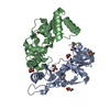
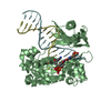
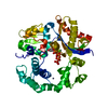
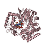
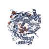
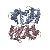
[English] 日本語
Yorodumi
- PDB-4by5: Crystal structure of Drosophila Frq2 -

+
Open data


ID or keywords:

Loading...

-
Basic information

Entry
Database: PDB / ID: 4by5
TitleCrystal structure of Drosophila Frq2
ComponentsFI18190P1
KeywordsCALCIUM-BINDING PROTEIN / CALCIUM SENSOR
Function / homology
Function and homology information


calcium sensitive guanylate cyclase activator activity / regulation of neurotransmitter secretion / neurotransmitter secretion / neuromuscular junction development / vesicle-mediated transport / synaptic vesicle / chemical synaptic transmission / calcium ion binding / cytoplasm
Similarity search - Function
Recoverin family / EF hand / EF-hand / Recoverin; domain 1 / EF-hand domain pair / EF-hand, calcium binding motif / EF-Hand 1, calcium-binding site / EF-hand calcium-binding domain. / EF-hand calcium-binding domain profile. / EF-hand domain ...Recoverin family / EF hand / EF-hand / Recoverin; domain 1 / EF-hand domain pair / EF-hand, calcium binding motif / EF-Hand 1, calcium-binding site / EF-hand calcium-binding domain. / EF-hand calcium-binding domain profile. / EF-hand domain / EF-hand domain pair / Orthogonal Bundle / Mainly Alpha
Similarity search - Domain/homology
Biological speciesDROSOPHILA MELANOGASTER (fruit fly)
MethodX-RAY DIFFRACTION / SYNCHROTRON / MOLECULAR REPLACEMENT / Resolution: 2.22 Å
AuthorsBanos-Mateos, S. / Chaves-Sanjuan, A. / Sanchez-Barrena, M.J.
CitationJournal: J.Cell.Sci. / Year: 2014
Title: The Guanine-Exchange Factor Ric8A Binds the Calcium Sensor Ncs-1 to Regulate Synapse Number and Probability of Release.
Authors: Romero-Pozuelo, J. / Dason, J.S. / Mansilla, A. / Banos-Mateos, S. / Sardina, J.L. / Chaves-Sanjuan, A. / Jurado-Gomez, J. / Santana, E. / Atwood, H.L. / Hernandez-Hernandez, A. / Sanchez- ...Authors: Romero-Pozuelo, J. / Dason, J.S. / Mansilla, A. / Banos-Mateos, S. / Sardina, J.L. / Chaves-Sanjuan, A. / Jurado-Gomez, J. / Santana, E. / Atwood, H.L. / Hernandez-Hernandez, A. / Sanchez-Barrena, M. / Ferrus, A.
History
DepositionJul 17, 2013Deposition site: PDBE / Processing site: PDBE
Revision 1.0Aug 13, 2014Provider: repository / Type: Initial release
Revision 1.1Dec 17, 2014Group: Database references
Revision 1.2Dec 20, 2023Group: Data collection / Database references ...Data collection / Database references / Derived calculations / Other / Refinement description
Category: chem_comp_atom / chem_comp_bond ...chem_comp_atom / chem_comp_bond / database_2 / pdbx_database_status / pdbx_initial_refinement_model / pdbx_struct_conn_angle / struct_conn / struct_ncs_dom_lim / struct_site
Item: _database_2.pdbx_DOI / _database_2.pdbx_database_accession ..._database_2.pdbx_DOI / _database_2.pdbx_database_accession / _pdbx_database_status.status_code_sf / _pdbx_struct_conn_angle.ptnr1_auth_comp_id / _pdbx_struct_conn_angle.ptnr1_auth_seq_id / _pdbx_struct_conn_angle.ptnr1_label_asym_id / _pdbx_struct_conn_angle.ptnr1_label_atom_id / _pdbx_struct_conn_angle.ptnr1_label_comp_id / _pdbx_struct_conn_angle.ptnr1_label_seq_id / _pdbx_struct_conn_angle.ptnr2_auth_comp_id / _pdbx_struct_conn_angle.ptnr2_auth_seq_id / _pdbx_struct_conn_angle.ptnr2_label_asym_id / _pdbx_struct_conn_angle.ptnr2_label_atom_id / _pdbx_struct_conn_angle.ptnr2_label_comp_id / _pdbx_struct_conn_angle.ptnr3_auth_comp_id / _pdbx_struct_conn_angle.ptnr3_auth_seq_id / _pdbx_struct_conn_angle.ptnr3_label_asym_id / _pdbx_struct_conn_angle.ptnr3_label_atom_id / _pdbx_struct_conn_angle.ptnr3_label_comp_id / _pdbx_struct_conn_angle.ptnr3_label_seq_id / _pdbx_struct_conn_angle.value / _struct_conn.pdbx_dist_value / _struct_conn.ptnr1_auth_comp_id / _struct_conn.ptnr1_auth_seq_id / _struct_conn.ptnr1_label_asym_id / _struct_conn.ptnr1_label_atom_id / _struct_conn.ptnr1_label_comp_id / _struct_conn.ptnr1_label_seq_id / _struct_conn.ptnr2_auth_comp_id / _struct_conn.ptnr2_auth_seq_id / _struct_conn.ptnr2_label_asym_id / _struct_conn.ptnr2_label_atom_id / _struct_conn.ptnr2_label_comp_id / _struct_conn.ptnr2_label_seq_id / _struct_ncs_dom_lim.beg_auth_comp_id / _struct_ncs_dom_lim.beg_label_asym_id / _struct_ncs_dom_lim.beg_label_comp_id / _struct_ncs_dom_lim.beg_label_seq_id / _struct_ncs_dom_lim.end_auth_comp_id / _struct_ncs_dom_lim.end_label_asym_id / _struct_ncs_dom_lim.end_label_comp_id / _struct_ncs_dom_lim.end_label_seq_id / _struct_site.pdbx_auth_asym_id / _struct_site.pdbx_auth_comp_id / _struct_site.pdbx_auth_seq_id

-
Structure visualization

Structure viewerMolecule:
MolmilJmol/JSmol

Downloads & links

-
Assembly

Deposited unit
A: FI18190P1
B: FI18190P1
C: FI18190P1
D: FI18190P1
hetero molecules


Theoretical massNumber of molelcules
Total (without water)88,19417
Polymers87,6904
Non-polymers50413
Water4,089227
1
A: FI18190P1
hetero molecules

C: FI18190P1
hetero molecules


Theoretical massNumber of molelcules
Total (without water)44,0858
Polymers43,8452
Non-polymers2406
Water362
TypeNameSymmetry operationNumber
identity operation1_555x,y,z1
crystal symmetry operation1_554x,y,z-11
Buried area2300 Å2
ΔGint-90.9 kcal/mol
Surface area18740 Å2
MethodPISA
2
C: FI18190P1
hetero molecules

A: FI18190P1
hetero molecules


Theoretical massNumber of molelcules
Total (without water)44,0858
Polymers43,8452
Non-polymers2406
Water362
TypeNameSymmetry operationNumber
identity operation1_555x,y,z1
crystal symmetry operation1_556x,y,z+11
Buried area2300 Å2
ΔGint-90.9 kcal/mol
Surface area18740 Å2
MethodPISA
3
B: FI18190P1
D: FI18190P1
hetero molecules


Theoretical massNumber of molelcules
Total (without water)44,1089
Polymers43,8452
Non-polymers2637
Water362
TypeNameSymmetry operationNumber
identity operation1_555x,y,z1
Buried area2460 Å2
ΔGint-99.4 kcal/mol
Surface area19090 Å2
MethodPISA
Unit cell
Length a, b, c (Å)55.984, 131.192, 56.710
Angle α, β, γ (deg.)90.00, 91.07, 90.00
Int Tables number4
Space group name H-MP1211
Noncrystallographic symmetry (NCS)NCS domain:
IDEns-IDDetails
11A
21B
12A
22C
13A
23D
14B
24C
15B
25D
16C
26D

NCS domain segments:

Component-ID: 0 / Refine code: 0

Dom-IDEns-IDBeg auth comp-IDBeg label comp-IDEnd auth comp-IDEnd label comp-IDAuth asym-IDLabel asym-IDAuth seq-IDLabel seq-ID
11ASNASNLEULEUAA5 - 1825 - 182
21ASNASNLEULEUBB5 - 1825 - 182
12LYSLYSALAALAAA7 - 1817 - 181
22LYSLYSALAALACC7 - 1817 - 181
13LYSLYSLEULEUAA7 - 1827 - 182
23LYSLYSLEULEUDD7 - 1827 - 182
14LYSLYSLEULEUBB7 - 1847 - 184
24LYSLYSLEULEUCC7 - 1847 - 184
15LYSLYSLEULEUBB7 - 1827 - 182
25LYSLYSLEULEUDD7 - 1827 - 182
16LYSLYSLEULEUCC7 - 1827 - 182
26LYSLYSLEULEUDD7 - 1827 - 182

NCS ensembles :
ID
1
2
3
4
5
6

-
Components

#1: Protein
FI18190P1 / FREQUENIN / FREQUENIN 2


Mass: 21922.443 Da / Num. of mol.: 4 / Mutation: YES
Source method: isolated from a genetically manipulated source
Details: CALCIUM BINDING TO EF HANDS 2,3 AND 4 / Source: (gene. exp.) DROSOPHILA MELANOGASTER (fruit fly) / Plasmid: PETDUET / Production host: ESCHERICHIA COLI (E. coli) / Strain (production host): BL21(DE3) / References: UniProt: Q9VWX8
#2: Chemical
ChemComp-CA / CALCIUM ION


Mass: 40.078 Da / Num. of mol.: 12 / Source method: obtained synthetically / Formula: Ca
#3: Chemical ChemComp-NA / SODIUM ION


Mass: 22.990 Da / Num. of mol.: 1 / Source method: obtained synthetically / Formula: Na
#4: Water ChemComp-HOH / water / Water


Mass: 18.015 Da / Num. of mol.: 227 / Source method: isolated from a natural source / Formula: H2O

-
Experimental details

-
Experiment

ExperimentMethod: X-RAY DIFFRACTION / Number of used crystals: 1

-
Sample preparation

CrystalDensity Matthews: 2.23 Å3/Da / Density % sol: 45.6 % / Description: NONE
Crystal growpH: 7.5 / Details: 21% PEG 4000, 0.1 M CACL2, 0.1 M HEPES PH 7.5

-
Data collection

DiffractionMean temperature: 100 K
Diffraction sourceSource: SYNCHROTRON / Site: ESRF / Beamline: ID14-1 / Wavelength: 0.9334
DetectorType: ADSC QUANTUM 210 / Detector: CCD / Date: Nov 5, 2011
RadiationProtocol: SINGLE WAVELENGTH / Monochromatic (M) / Laue (L): M / Scattering type: x-ray
Radiation wavelengthWavelength: 0.9334 Å / Relative weight: 1
ReflectionResolution: 2.22→42.97 Å / Num. obs: 40192 / % possible obs: 99.4 % / Observed criterion σ(I): 2 / Redundancy: 3.7 % / Rmerge(I) obs: 0.08 / Net I/σ(I): 12.2
Reflection shellResolution: 2.22→2.34 Å / Redundancy: 3.3 % / Rmerge(I) obs: 0.47 / Mean I/σ(I) obs: 2.6 / % possible all: 96.5

-
Processing

Software
NameVersionClassification
REFMAC5.7.0029refinement
PHASERphasing
RefinementMethod to determine structure: MOLECULAR REPLACEMENT
Starting model: PDB ENTRY 1G8I
Resolution: 2.22→42.93 Å / Cor.coef. Fo:Fc: 0.936 / Cor.coef. Fo:Fc free: 0.89 / SU B: 6.955 / SU ML: 0.174 / Cross valid method: THROUGHOUT / ESU R: 0.307 / ESU R Free: 0.242 / Stereochemistry target values: MAXIMUM LIKELIHOOD / Details: HYDROGENS HAVE BEEN ADDED IN THE RIDING POSITIONS.
RfactorNum. reflection% reflectionSelection details
Rfree0.27052 2009 5 %RANDOM
Rwork0.21068 ---
obs0.21366 38151 99.64 %-
Solvent computationIon probe radii: 0.8 Å / Shrinkage radii: 0.8 Å / VDW probe radii: 1.2 Å / Solvent model: MASK
Displacement parametersBiso mean: 39.466 Å2
Baniso -1Baniso -2Baniso -3
1--0.45 Å20 Å2-0.06 Å2
2--2.37 Å20 Å2
3----1.92 Å2
Refinement stepCycle: LAST / Resolution: 2.22→42.93 Å
ProteinNucleic acidLigandSolventTotal
Num. atoms5702 0 13 227 5942
Refine LS restraints
Refine-IDTypeDev idealDev ideal targetNumber
X-RAY DIFFRACTIONr_bond_refined_d0.0160.0195828
X-RAY DIFFRACTIONr_bond_other_d
X-RAY DIFFRACTIONr_angle_refined_deg1.7921.9477840
X-RAY DIFFRACTIONr_angle_other_deg
X-RAY DIFFRACTIONr_dihedral_angle_1_deg5.8855680
X-RAY DIFFRACTIONr_dihedral_angle_2_deg35.25224.543339
X-RAY DIFFRACTIONr_dihedral_angle_3_deg21.761151067
X-RAY DIFFRACTIONr_dihedral_angle_4_deg21.2831545
X-RAY DIFFRACTIONr_chiral_restr0.1240.2817
X-RAY DIFFRACTIONr_gen_planes_refined0.0090.0214517
X-RAY DIFFRACTIONr_gen_planes_other
X-RAY DIFFRACTIONr_nbd_refined
X-RAY DIFFRACTIONr_nbd_other
X-RAY DIFFRACTIONr_nbtor_refined
X-RAY DIFFRACTIONr_nbtor_other
X-RAY DIFFRACTIONr_xyhbond_nbd_refined
X-RAY DIFFRACTIONr_xyhbond_nbd_other
X-RAY DIFFRACTIONr_metal_ion_refined
X-RAY DIFFRACTIONr_metal_ion_other
X-RAY DIFFRACTIONr_symmetry_vdw_refined
X-RAY DIFFRACTIONr_symmetry_vdw_other
X-RAY DIFFRACTIONr_symmetry_hbond_refined
X-RAY DIFFRACTIONr_symmetry_hbond_other
X-RAY DIFFRACTIONr_symmetry_metal_ion_refined
X-RAY DIFFRACTIONr_symmetry_metal_ion_other
X-RAY DIFFRACTIONr_mcbond_it
X-RAY DIFFRACTIONr_mcbond_other
X-RAY DIFFRACTIONr_mcangle_it
X-RAY DIFFRACTIONr_mcangle_other
X-RAY DIFFRACTIONr_scbond_it
X-RAY DIFFRACTIONr_scbond_other
X-RAY DIFFRACTIONr_scangle_it
X-RAY DIFFRACTIONr_scangle_other
X-RAY DIFFRACTIONr_long_range_B_refined
X-RAY DIFFRACTIONr_long_range_B_other
X-RAY DIFFRACTIONr_rigid_bond_restr
X-RAY DIFFRACTIONr_sphericity_free
X-RAY DIFFRACTIONr_sphericity_bonded
Refine LS restraints NCS

Refine-ID: X-RAY DIFFRACTION / Type: interatomic distance / Weight position: 0.05

Ens-IDDom-IDAuth asym-IDNumberRms dev position (Å)
11A2410.17
12B2410.17
21A2230.21
22C2230.21
31A2250.26
32D2250.26
41B2190.24
42C2190.24
51B2130.27
52D2130.27
61C2200.26
62D2200.26
LS refinement shellResolution: 2.221→2.279 Å / Total num. of bins used: 20
RfactorNum. reflection% reflection
Rfree0.332 124 -
Rwork0.263 2726 -
obs--96.28 %

+
About Yorodumi

-
News

-
Feb 9, 2022. New format data for meta-information of EMDB entries

New format data for meta-information of EMDB entries

  • Version 3 of the EMDB header file is now the official format.
  • The previous official version 1.9 will be removed from the archive.

Related info.:EMDB header

External links:wwPDB to switch to version 3 of the EMDB data model

-
Aug 12, 2020. Covid-19 info

Covid-19 info

URL: https://pdbj.org/emnavi/covid19.php

New page: Covid-19 featured information page in EM Navigator.

Related info.:Covid-19 info / Mar 5, 2020. Novel coronavirus structure data

+
Mar 5, 2020. Novel coronavirus structure data

Novel coronavirus structure data

Related info.:Yorodumi Speices / Aug 12, 2020. Covid-19 info

External links:COVID-19 featured content - PDBj / Molecule of the Month (242):Coronavirus Proteases

+
Jan 31, 2019. EMDB accession codes are about to change! (news from PDBe EMDB page)

EMDB accession codes are about to change! (news from PDBe EMDB page)

  • The allocation of 4 digits for EMDB accession codes will soon come to an end. Whilst these codes will remain in use, new EMDB accession codes will include an additional digit and will expand incrementally as the available range of codes is exhausted. The current 4-digit format prefixed with “EMD-” (i.e. EMD-XXXX) will advance to a 5-digit format (i.e. EMD-XXXXX), and so on. It is currently estimated that the 4-digit codes will be depleted around Spring 2019, at which point the 5-digit format will come into force.
  • The EM Navigator/Yorodumi systems omit the EMD- prefix.

Related info.:Q: What is EMD? / ID/Accession-code notation in Yorodumi/EM Navigator

External links:EMDB Accession Codes are Changing Soon! / Contact to PDBj

+
Jul 12, 2017. Major update of PDB

Major update of PDB

  • wwPDB released updated PDB data conforming to the new PDBx/mmCIF dictionary.
  • This is a major update changing the version number from 4 to 5, and with Remediation, in which all the entries are updated.
  • In this update, many items about electron microscopy experimental information are reorganized (e.g. em_software).
  • Now, EM Navigator and Yorodumi are based on the updated data.

External links:wwPDB Remediation / Enriched Model Files Conforming to OneDep Data Standards Now Available in the PDB FTP Archive

-
Yorodumi

Thousand views of thousand structures

  • Yorodumi is a browser for structure data from EMDB, PDB, SASBDB, etc.
  • This page is also the successor to EM Navigator detail page, and also detail information page/front-end page for Omokage search.
  • The word "yorodu" (or yorozu) is an old Japanese word meaning "ten thousand". "mi" (miru) is to see.

Related info.:EMDB / PDB / SASBDB / Comparison of 3 databanks / Yorodumi Search / Aug 31, 2016. New EM Navigator & Yorodumi / Yorodumi Papers / Jmol/JSmol / Function and homology information / Changes in new EM Navigator and Yorodumi

Read more