 Movie
Movie Controller

[English] 日本語
 Yorodumi
- PDB-4bx0: Crystal Structure of a Monomeric Variant of murine Chronophin (Py... -
+ Open data

- Basic information
| Entry | Database: PDB / ID: 4bx0 | |||||||||
|---|---|---|---|---|---|---|---|---|---|---|
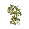
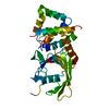
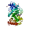
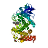
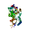
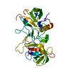
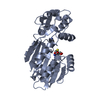
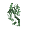
| Title | Crystal Structure of a Monomeric Variant of murine Chronophin (Pyridoxal Phosphate phosphatase) | |||||||||
 Components Components | PYRIDOXAL PHOSPHATE PHOSPHATASE | |||||||||
 Keywords Keywords |  HYDROLASE / PDXP / PLPP / HAD PHOSPHATASE / HAD-LIKE HYDROLASE / MONOMER HYDROLASE / PDXP / PLPP / HAD PHOSPHATASE / HAD-LIKE HYDROLASE / MONOMER | |||||||||
| Function / homology |  Function and homology informationpyridoxal phosphatase / pyridoxal phosphate catabolic process / actin rod assembly / pyridoxal phosphatase activity / contractile ring / positive regulation of actin filament depolymerization / cellular response to ATP / regulation of mitotic nuclear division / myosin phosphatase activity / protein-serine/threonine phosphatase ...pyridoxal phosphatase / pyridoxal phosphate catabolic process / actin rod assembly / pyridoxal phosphatase activity / contractile ring / positive regulation of actin filament depolymerization / cellular response to ATP / regulation of mitotic nuclear division / myosin phosphatase activity / protein-serine/threonine phosphatase / growth factor binding / phosphoprotein phosphatase activity / cleavage furrow / lamellipodium membrane / dephosphorylation / heat shock protein binding / protein dephosphorylation / regulation of cytokinesis / ruffle membrane / cell-cell junction / actin cytoskeleton / midbody / magnesium ion binding / protein homodimerization activity / cytosol / cytoplasm Function and homology informationpyridoxal phosphatase / pyridoxal phosphate catabolic process / actin rod assembly / pyridoxal phosphatase activity / contractile ring / positive regulation of actin filament depolymerization / cellular response to ATP / regulation of mitotic nuclear division / myosin phosphatase activity / protein-serine/threonine phosphatase ...pyridoxal phosphatase / pyridoxal phosphate catabolic process / actin rod assembly / pyridoxal phosphatase activity / contractile ring / positive regulation of actin filament depolymerization / cellular response to ATP / regulation of mitotic nuclear division / myosin phosphatase activity / protein-serine/threonine phosphatase / growth factor binding / phosphoprotein phosphatase activity / cleavage furrow / lamellipodium membrane / dephosphorylation / heat shock protein binding / protein dephosphorylation / regulation of cytokinesis / ruffle membrane / cell-cell junction / actin cytoskeleton / midbody / magnesium ion binding / protein homodimerization activity / cytosol / cytoplasmSimilarity search - Function | |||||||||
| Biological species |  MUS MUSCULUS (house mouse) MUS MUSCULUS (house mouse) | |||||||||
| Method | X-RAY DIFFRACTION / SYNCHROTRON / MOLECULAR REPLACEMENT / Resolution: 1.75 Å | |||||||||
 Authors Authors | Kestler, C. / Knobloch, G. / Gohla, A. / Schindelin, H. | |||||||||
 Citation Citation | Journal: J.Biol.Chem. / Year: 2014 Title: Chronophin Dimerization is Required for Proper Positioning of its Substrate Specificity Loop Authors: Kestler, C. / Knobloch, G. / Tessmer, I. / Jeanclos, E. / Schindelin, H. / Gohla, A. | |||||||||
| History |
|
- Structure visualization
| Structure viewer | Molecule: MolmilJmol/JSmol |
|---|
- Downloads & links
-Download
| PDBx/mmCIF format | 4bx0.cif.gz | 179 KB | Display | PDBx/mmCIF format |
|---|---|---|---|---|
| PDB format | pdb4bx0.ent.gz | 144.1 KB | Display | PDB format |
| PDBx/mmJSON format | 4bx0.json.gz | Tree view | PDBx/mmJSON format | |
| Others |  Other downloads Other downloads |
-Validation report
| Arichive directory | https://data.pdbj.org/pub/pdb/validation_reports/bx/4bx0ftp://data.pdbj.org/pub/pdb/validation_reports/bx/4bx0 | HTTPS FTP |
|---|
-Related structure data
| Related structure data |  4bx2C 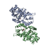 4bx3C 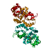 2oycS C: citing same article ( S: Starting model for refinement |
|---|---|
| Similar structure data |


-Links
 PDBj
PDBj- Assembly
| Deposited unit | 
| ||||||||
|---|---|---|---|---|---|---|---|---|---|
| 1 |
| ||||||||
| Unit cell |
|
-Components
| #1: Protein | Mass: 31664.041 Da / Num. of mol.: 1 / Mutation: YES Source method: isolated from a genetically manipulated source Source: (gene. exp.) MUS MUSCULUS (house mouse) / Plasmid: PETM-11 / Production host:  ESCHERICHIA COLI (E. coli) / Strain (production host): BL21(DE3) ESCHERICHIA COLI (E. coli) / Strain (production host): BL21(DE3)References: UniProt: P60487, pyridoxal phosphatase, phosphoserine phosphatase |
|---|---|
| #2: Chemical | ChemComp-MG /   Mass: 24.305 Da / Num. of mol.: 1 / Source method: obtained synthetically / Formula: Mg Mass: 24.305 Da / Num. of mol.: 1 / Source method: obtained synthetically / Formula: Mg |
| #3: Chemical | ChemComp-GOL / Glycerol  Mass: 92.094 Da / Num. of mol.: 1 / Source method: obtained synthetically / Formula: C3H8O3 Mass: 92.094 Da / Num. of mol.: 1 / Source method: obtained synthetically / Formula: C3H8O3 |
| #4: Water | ChemComp-HOH / Water Mass: 18.015 Da / Num. of mol.: 242 / Source method: isolated from a natural source / Formula: H2O Mass: 18.015 Da / Num. of mol.: 242 / Source method: isolated from a natural source / Formula: H2O |
-Experimental details
-Experiment
| Experiment | Method: X-RAY DIFFRACTION / Number of used crystals: 1 |
|---|
- Sample preparation
| Crystal | Density Matthews: 2.01 Å3/Da / Density % sol: 38.85 % / Description: NONE |
|---|---|
| Crystal grow | pH: 6.5 / Details: 0.1M MES PH 6.5, 25% PEG-MME 550 |
-Data collection
| Diffraction | Mean temperature: 100 K |
|---|---|
| Diffraction source | Source: SYNCHROTRON / Site: BESSY  / Beamline: 14.1 / Wavelength: 0.91841 / Beamline: 14.1 / Wavelength: 0.91841 |
| Detector | Type: MARMOSAIC 225 mm CCD / Detector: CCD / Date: Apr 17, 2012 / Details: MIRRORS |
| Radiation | Protocol: SINGLE WAVELENGTH / Monochromatic (M) / Laue (L): M / Scattering type: x-ray |
| Radiation wavelength | Wavelength: 0.91841 Å / Relative weight: 1 |
| Reflection | Resolution: 1.75→30.61 Å / Num. obs: 25533 / % possible obs: 99.8 % / Observed criterion σ(I): -3 / Redundancy: 3.5 % / Biso Wilson estimate: 19.09 Å2 / Rmerge(I) obs: 0.08 / Net I/σ(I): 10.2 |
| Reflection shell | Resolution: 1.75→1.84 Å / Redundancy: 3.5 % / Rmerge(I) obs: 0.56 / Mean I/σ(I) obs: 2.3 / % possible all: 99.9 |
- Processing
| Software |
| ||||||||||||||||||||||||||||||||||||||||||||||||||||||||||||||||||||||
|---|---|---|---|---|---|---|---|---|---|---|---|---|---|---|---|---|---|---|---|---|---|---|---|---|---|---|---|---|---|---|---|---|---|---|---|---|---|---|---|---|---|---|---|---|---|---|---|---|---|---|---|---|---|---|---|---|---|---|---|---|---|---|---|---|---|---|---|---|---|---|---|
| Refinement | Method to determine structure: MOLECULAR REPLACEMENT Starting model: PDB ENTRY 2OYC Resolution: 1.75→28.404 Å / SU ML: 0.28 / σ(F): 1.37 / Phase error: 23.44 / Stereochemistry target values: ML
| ||||||||||||||||||||||||||||||||||||||||||||||||||||||||||||||||||||||
| Solvent computation | Shrinkage radii: 0.86 Å / VDW probe radii: 1.1 Å / Solvent model: FLAT BULK SOLVENT MODEL / Bsol: 43.901 Å2 / ksol: 0.353 e/Å3 | ||||||||||||||||||||||||||||||||||||||||||||||||||||||||||||||||||||||
| Displacement parameters | Biso mean: 28.6 Å2
| ||||||||||||||||||||||||||||||||||||||||||||||||||||||||||||||||||||||
| Refinement step | Cycle: LAST / Resolution: 1.75→28.404 Å
| ||||||||||||||||||||||||||||||||||||||||||||||||||||||||||||||||||||||
| Refine LS restraints |
| ||||||||||||||||||||||||||||||||||||||||||||||||||||||||||||||||||||||
| LS refinement shell |
|
