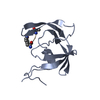
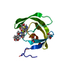
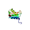
Entry Database : PDB / ID : 4bgjTitle Crystal structure of the phox-homology domain of human sorting nexin 14 SORTING NEXIN-14 Keywords Function / homology Function Domain/homology Component
/ / / / / / / / / / / / / / / / / / / / / / / / / / / / / / / / / / / / / / / / / / / / / / / / / / / / Biological species HOMO SAPIENS (human)Method / / / Resolution : 2.55 Å Authors Vollmar, M. / Kiyani, W. / Shrestha, L. / Goubin, S. / Krojer, T. / Pike, A.C.W. / Carpenter, E. / Quigley, A. / McKenzie, A. / Burgess-Brown, N. ...Vollmar, M. / Kiyani, W. / Shrestha, L. / Goubin, S. / Krojer, T. / Pike, A.C.W. / Carpenter, E. / Quigley, A. / McKenzie, A. / Burgess-Brown, N. / von Delft, F. / Arrowsmith, C.H. / Bountra, C. / Edwards, A. / Yue, W.W. Journal : To be Published Title : Crystal Structure of the Phox-Homology Domain of Human Sorting Nexin 14Authors: Vollmar, M. / Kiyani, W. / Shrestha, L. / Goubin, S. / Krojer, T. / Pike, A.C.W. / Carpenter, E. / Quigley, A. / Mckenzie, A. / Burgess-Brown, N. / von Delft, F. / Arrowsmith, C.H. / ... Authors : Vollmar, M. / Kiyani, W. / Shrestha, L. / Goubin, S. / Krojer, T. / Pike, A.C.W. / Carpenter, E. / Quigley, A. / Mckenzie, A. / Burgess-Brown, N. / von Delft, F. / Arrowsmith, C.H. / Bountra, C. / Edwards, A. / Yue, W.W. History Deposition Mar 27, 2013 Deposition site / Processing site Revision 1.0 May 29, 2013 Provider / Type Revision 1.1 Jan 24, 2018 Group / Category / Item Revision 1.2 Dec 20, 2023 Group Data collection / Database references ... Data collection / Database references / Other / Refinement description Category chem_comp_atom / chem_comp_bond ... chem_comp_atom / chem_comp_bond / database_2 / pdbx_database_status / pdbx_initial_refinement_model Item / _database_2.pdbx_database_accession / _pdbx_database_status.status_code_sf
Show all Show less
 Movie
Movie Controller

 Yorodumi
 Open data
 Basic information
 Components
 Keywords
 Function and homology information
 Authors
 Citation
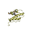
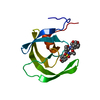
 Structure visualization
 Downloads & links
 Other downloads

 PDBj
PDBj
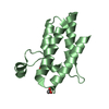
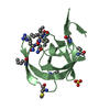
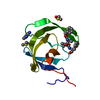
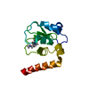
 Assembly
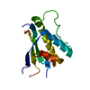

 Mass: 18.015 Da / Num. of mol.: 4 / Source method: isolated from a natural source / Formula: H2O
 Sample preparation
 / Beamline: I04 / Wavelength: 1.0163
  Processing