 Movie
Movie Controller


+ Open data

- Basic information
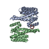
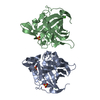
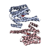
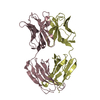
| Entry | Database: PDB / ID: 4bg6 | ||||||
|---|---|---|---|---|---|---|---|
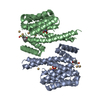
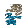
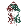
| Title | 14-3-3 interaction with Rnd3 prenyl-phosphorylation motif | ||||||
 Components Components |
| ||||||
 Keywords Keywords |  SIGNALING PROTEIN / PRENYLATION / ACTIN CYTOSKELETON SIGNALING PROTEIN / PRENYLATION / ACTIN CYTOSKELETON | ||||||
| Function / homology |  Function and homology information Function and homology informationGolgi reassembly / regulation of synapse maturation / NOTCH4 Activation and Transmission of Signal to the Nucleus / establishment of Golgi localization / Rap1 signalling / negative regulation of protein localization to nucleus / RND3 GTPase cycle / KSRP (KHSRP) binds and destabilizes mRNA / small GTPase-mediated signal transduction / GP1b-IX-V activation signalling ...Golgi reassembly / regulation of synapse maturation / NOTCH4 Activation and Transmission of Signal to the Nucleus / establishment of Golgi localization / Rap1 signalling / negative regulation of protein localization to nucleus / RND3 GTPase cycle / KSRP (KHSRP) binds and destabilizes mRNA / small GTPase-mediated signal transduction / GP1b-IX-V activation signalling / Regulation of localization of FOXO transcription factors / Interleukin-3, Interleukin-5 and GM-CSF signaling / phosphoserine residue binding / Activation of BAD and translocation to mitochondria / SARS-CoV-2 targets host intracellular signalling and regulatory pathways / cellular response to glucose starvation / Chk1/Chk2(Cds1) mediated inactivation of Cyclin B:Cdk1 complex / SARS-CoV-1 targets host intracellular signalling and regulatory pathways / RHO GTPases activate PKNs / negative regulation of TORC1 signaling / negative regulation of innate immune response / regulation of ERK1 and ERK2 cascade / protein sequestering activity / actin filament organization / Translocation of SLC2A4 (GLUT4) to the plasma membrane / Deactivation of the beta-catenin transactivating complex / TP53 Regulates Metabolic Genes / Negative regulation of NOTCH4 signaling / cell migration / melanosome / actin cytoskeleton organization / blood microparticle / DNA-binding transcription factor binding / vesicle / transmembrane transporter binding / cell adhesion / cadherin binding / Golgi membrane / protein phosphorylation / focal adhesion / GTPase activity / glutamatergic synapse / ubiquitin protein ligase binding / GTP binding / negative regulation of apoptotic process / protein kinase binding / negative regulation of transcription by RNA polymerase II / signal transduction / extracellular space / RNA binding / extracellular exosome / nucleoplasm / identical protein binding / nucleus / plasma membrane / cytosol / cytoplasmSimilarity search - Function | ||||||
| Biological species |  HOMO SAPIENS (human) HOMO SAPIENS (human) | ||||||
| Method | X-RAY DIFFRACTION / SYNCHROTRON / MOLECULAR REPLACEMENT / Resolution: 2.3 Å | ||||||
 Authors Authors | Riou, P. / Kjaer, S. / Purkiss, A. / O'Reilly, N. / McDonald, N.Q. | ||||||
 Citation Citation | Journal: Cell(Cambridge,Mass.) / Year: 2013 Title: 14-3-3 Proteins Interact with a Hybrid Prenyl-Phosphorylation Motif to Inhibit G Proteins. Authors: Riou, P. / Kjaer, S. / Garg, R. / Purkiss, A. / George, R. / Cain, R.J. / Bineva, G. / Reymond, N. / Mccoll, B. / Thompson, A.J. / O'Reilly, N. / Mcdonald, N.Q. / Parker, P.J. / Ridley, A.J. | ||||||
| History |
|
- Structure visualization
| Structure viewer | Molecule: MolmilJmol/JSmol |
|---|
- Downloads & links
-Download
| PDBx/mmCIF format | 4bg6.cif.gz | 199.9 KB | Display | PDBx/mmCIF format |
|---|---|---|---|---|
| PDB format | pdb4bg6.ent.gz | 159.1 KB | Display | PDB format |
| PDBx/mmJSON format | 4bg6.json.gz | Tree view | PDBx/mmJSON format | |
| Others |  Other downloads Other downloads |
-Validation report
| Arichive directory | https://data.pdbj.org/pub/pdb/validation_reports/bg/4bg6ftp://data.pdbj.org/pub/pdb/validation_reports/bg/4bg6 | HTTPS FTP |
|---|
-Related structure data
| Related structure data |  1wh0S S: Starting model for refinement |
|---|---|
| Similar structure data |


-Links
 PDBj
PDBj


- Assembly
| Deposited unit | 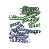
| ||||||||
|---|---|---|---|---|---|---|---|---|---|
| 1 | 
| ||||||||
| Unit cell |
| ||||||||
| Noncrystallographic symmetry (NCS) | NCS oper: (Code: given Matrix: (-0.99735, -0.07218, -0.00942), Vector : |
-Components
| #1: Protein | Mass: 27777.092 Da / Num. of mol.: 2 Source method: isolated from a genetically manipulated source Source: (gene. exp.) HOMO SAPIENS (human) / Production host:  ESCHERICHIA COLI (E. coli) / Strain (production host): BL21(DE3) / Variant (production host): STAR / References: UniProt: P63104 ESCHERICHIA COLI (E. coli) / Strain (production host): BL21(DE3) / Variant (production host): STAR / References: UniProt: P63104#2: Protein/peptide | Mass: 1247.359 Da / Num. of mol.: 2 / Fragment: C-TERMINUS, RESIDUES 232-241 / Source method: obtained synthetically / Details: FARNESYLATION ON CYS 347 / Source: (synth.) HOMO SAPIENS (human) / References: UniProt: P61587#3: Chemical | Polyethylene glycol  Mass: 354.436 Da / Num. of mol.: 3 / Source method: obtained synthetically / Formula: C16H34O8 / Comment: precipitant*YM Mass: 354.436 Da / Num. of mol.: 3 / Source method: obtained synthetically / Formula: C16H34O8 / Comment: precipitant*YM#4: Chemical | Farnesol  Mass: 206.367 Da / Num. of mol.: 2 / Source method: obtained synthetically / Formula: C15H26 Mass: 206.367 Da / Num. of mol.: 2 / Source method: obtained synthetically / Formula: C15H26#5: Water | ChemComp-HOH / | Water Mass: 18.015 Da / Num. of mol.: 80 / Source method: isolated from a natural source / Formula: H2O Mass: 18.015 Da / Num. of mol.: 80 / Source method: isolated from a natural source / Formula: H2O |
|---|
-Experimental details
-Experiment
| Experiment | Method: X-RAY DIFFRACTION / Number of used crystals: 1 |
|---|
- Sample preparation
| Crystal | Density Matthews: 3.08 Å3/Da / Density % sol: 60.02 % / Description: NONE |
|---|---|
| Crystal grow | Method: vapor diffusion, sitting drop / pH: 8.5 Details: 0.17 M SODIUM ACETATE; 0.085 M TRIS, PH 8.5; 25.5% (W/V) PEG4000, 15% (V/V) GLYCEROL |
-Data collection
| Diffraction | Mean temperature: 100 K |
|---|---|
| Diffraction source | Source: SYNCHROTRON / Site: Diamond  / Beamline: I24 / Wavelength: 0.9763 / Beamline: I24 / Wavelength: 0.9763 |
| Detector | Type: DECTRIS PILATUS 6M / Detector: PIXEL / Date: Apr 18, 2011 / Details: MIRRORS |
| Radiation | Monochromator: SI CRYSTAL / Protocol: SINGLE WAVELENGTH / Monochromatic (M) / Laue (L): M / Scattering type: x-ray |
| Radiation wavelength | Wavelength: 0.9763 Å / Relative weight: 1 |
| Reflection | Resolution: 2.3→38.51 Å / Num. obs: 28918 / % possible obs: 99.1 % / Observed criterion σ(I): 5 / Redundancy: 3.21 % / Biso Wilson estimate: 56.28 Å2 / Rmerge(I) obs: 0.08 / Net I/σ(I): 5.9 |
| Reflection shell | Resolution: 2.3→2.38 Å / Redundancy: 3.2 % / Rmerge(I) obs: 0.54 / Mean I/σ(I) obs: 1.2 / % possible all: 99.1 |
- Processing
| Software |
| ||||||||||||||||||||||||||||||||||||||||||||||||||||||||||||||||||||||||||||||||||||||||||||||||||||||||||||||||||||||||||||||||||||||||||||||||||||||||||||||||||||||||||||||||||||||||||||||||||||||||
|---|---|---|---|---|---|---|---|---|---|---|---|---|---|---|---|---|---|---|---|---|---|---|---|---|---|---|---|---|---|---|---|---|---|---|---|---|---|---|---|---|---|---|---|---|---|---|---|---|---|---|---|---|---|---|---|---|---|---|---|---|---|---|---|---|---|---|---|---|---|---|---|---|---|---|---|---|---|---|---|---|---|---|---|---|---|---|---|---|---|---|---|---|---|---|---|---|---|---|---|---|---|---|---|---|---|---|---|---|---|---|---|---|---|---|---|---|---|---|---|---|---|---|---|---|---|---|---|---|---|---|---|---|---|---|---|---|---|---|---|---|---|---|---|---|---|---|---|---|---|---|---|---|---|---|---|---|---|---|---|---|---|---|---|---|---|---|---|---|---|---|---|---|---|---|---|---|---|---|---|---|---|---|---|---|---|---|---|---|---|---|---|---|---|---|---|---|---|---|---|---|---|
| Refinement | Method to determine structure: MOLECULAR REPLACEMENT Starting model: PDB ENTRY 1WH0 Resolution: 2.3→38.508 Å / SU ML: 0.41 / σ(F): 1.34 / Phase error: 35.7 / Stereochemistry target values: ML
| ||||||||||||||||||||||||||||||||||||||||||||||||||||||||||||||||||||||||||||||||||||||||||||||||||||||||||||||||||||||||||||||||||||||||||||||||||||||||||||||||||||||||||||||||||||||||||||||||||||||||
| Solvent computation | Shrinkage radii: 1 Å / VDW probe radii: 1.2 Å / Solvent model: FLAT BULK SOLVENT MODEL | ||||||||||||||||||||||||||||||||||||||||||||||||||||||||||||||||||||||||||||||||||||||||||||||||||||||||||||||||||||||||||||||||||||||||||||||||||||||||||||||||||||||||||||||||||||||||||||||||||||||||
| Displacement parameters | Biso mean: 69.018 Å2 | ||||||||||||||||||||||||||||||||||||||||||||||||||||||||||||||||||||||||||||||||||||||||||||||||||||||||||||||||||||||||||||||||||||||||||||||||||||||||||||||||||||||||||||||||||||||||||||||||||||||||
| Refinement step | Cycle: LAST / Resolution: 2.3→38.508 Å
| ||||||||||||||||||||||||||||||||||||||||||||||||||||||||||||||||||||||||||||||||||||||||||||||||||||||||||||||||||||||||||||||||||||||||||||||||||||||||||||||||||||||||||||||||||||||||||||||||||||||||
| Refine LS restraints |
| ||||||||||||||||||||||||||||||||||||||||||||||||||||||||||||||||||||||||||||||||||||||||||||||||||||||||||||||||||||||||||||||||||||||||||||||||||||||||||||||||||||||||||||||||||||||||||||||||||||||||
| LS refinement shell |
| ||||||||||||||||||||||||||||||||||||||||||||||||||||||||||||||||||||||||||||||||||||||||||||||||||||||||||||||||||||||||||||||||||||||||||||||||||||||||||||||||||||||||||||||||||||||||||||||||||||||||
| Refinement TLS params. | Method: refined / Refine-ID: X-RAY DIFFRACTION
| ||||||||||||||||||||||||||||||||||||||||||||||||||||||||||||||||||||||||||||||||||||||||||||||||||||||||||||||||||||||||||||||||||||||||||||||||||||||||||||||||||||||||||||||||||||||||||||||||||||||||
| Refinement TLS group |
|
