 Movie
Movie Controller

[English] 日本語
 Yorodumi
- PDB-4b6j: Crystal structure of phosphoserine phosphatase from T. onnurineus -
+ Open data

- Basic information
| Entry | Database: PDB / ID: 4b6j | ||||||
|---|---|---|---|---|---|---|---|
| Title | Crystal structure of phosphoserine phosphatase from T. onnurineus | ||||||
 Components Components | PHOSPHOSERINE PHOSPHATASE | ||||||
 Keywords Keywords | HYDROLASE / L-SERINE / HALOACID DEHALOGENASE SUPERFAMILY | ||||||
| Function / homology |  Function and homology information Function and homology information | ||||||
| Biological species |   THERMOCOCCUS ONNURINEUS NA1 (archaea) THERMOCOCCUS ONNURINEUS NA1 (archaea) | ||||||
| Method | X-RAY DIFFRACTION / MOLECULAR REPLACEMENT / Resolution: 3.34 Å | ||||||
 Authors Authors | Jung, T.-Y. / Kim, Y.-S. / Song, H.-N. / Woo, E. | ||||||
 Citation Citation | Journal: Proteins / Year: 2013 Title: Identification of a Novel Ligand Binding Site in Phosphoserine Phosphatase from the Hyperthermophilic Archaeon Thermococcus Onnurineus. Authors: Jung, T.-Y. / S-Kim, Y. / Oh, B.H. / Woo, E. | ||||||
| History |
|
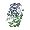
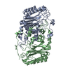
- Structure visualization
| Structure viewer | Molecule: MolmilJmol/JSmol |
|---|
- Downloads & links
-Download
| PDBx/mmCIF format | 4b6j.cif.gz | 164.4 KB | Display | PDBx/mmCIF format |
|---|---|---|---|---|
| PDB format | pdb4b6j.ent.gz | 132.8 KB | Display | PDB format |
| PDBx/mmJSON format | 4b6j.json.gz | Tree view | PDBx/mmJSON format | |
| Others |  Other downloads Other downloads |
-Validation report
| Arichive directory | https://data.pdbj.org/pub/pdb/validation_reports/b6/4b6jftp://data.pdbj.org/pub/pdb/validation_reports/b6/4b6j | HTTPS FTP |
|---|
-Related structure data
| Related structure data |  4ap9SC S: Starting model for refinement C: citing same article ( |
|---|---|
| Similar structure data |


-Links
 PDBj
PDBj

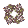
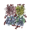
- Assembly
| Deposited unit | 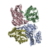
| ||||||||||||||||||
|---|---|---|---|---|---|---|---|---|---|---|---|---|---|---|---|---|---|---|---|
| 1 | 
| ||||||||||||||||||
| Unit cell |
| ||||||||||||||||||
| Noncrystallographic symmetry (NCS) | NCS domain:
NCS domain segments:
|
-Components
| #1: Protein | Mass: 23018.645 Da / Num. of mol.: 4 Source method: isolated from a genetically manipulated source Source: (gene. exp.) THERMOCOCCUS ONNURINEUS NA1 (archaea) / Production host:  ESCHERICHIA COLI MC1061 (bacteria) / Variant (production host): ARAD139, GALE15, GALK16 / References: UniProt: B6YX36, phosphoserine phosphatase ESCHERICHIA COLI MC1061 (bacteria) / Variant (production host): ARAD139, GALE15, GALK16 / References: UniProt: B6YX36, phosphoserine phosphatase |
|---|
-Experimental details
-Experiment
| Experiment | Method: X-RAY DIFFRACTION / Number of used crystals: 1 |
|---|
- Sample preparation
| Crystal | Density Matthews: 2.61 Å3/Da / Density % sol: 52.9 % / Description: NONE |
|---|---|
| Crystal grow | pH: 6.5 Details: 30% PENTAERYTHRITOL ETHOXYLATE, 50 MM BIS-TRIS PH 6.5, 50 MM AMMONIUM SULFATE |
-Data collection
| Diffraction | Mean temperature: 95 K |
|---|---|
| Diffraction source | Source: ROTATING ANODE / Type: RIGAKU / Wavelength: 1.5418 |
| Detector | Type: RIGAKU R-AXIS IV / Detector: IMAGE PLATE / Date: Sep 29, 2011 |
| Radiation | Protocol: SINGLE WAVELENGTH / Monochromatic (M) / Laue (L): M / Scattering type: x-ray |
| Radiation wavelength | Wavelength: 1.5418 Å / Relative weight: 1 |
| Reflection | Resolution: 3.34→44.64 Å / Num. obs: 14085 / % possible obs: 99.6 % / Observed criterion σ(I): 0 / Redundancy: 3.1 % / Biso Wilson estimate: 66.18 Å2 / Rmerge(I) obs: 0.18 / Net I/σ(I): 9.1 |
| Reflection shell | Resolution: 3.34→3.4 Å / Redundancy: 2.9 % / Rmerge(I) obs: 0.54 / Mean I/σ(I) obs: 2.2 / % possible all: 99.6 |
- Processing
| Software |
| ||||||||||||||||||||||||||||||||||||||||||
|---|---|---|---|---|---|---|---|---|---|---|---|---|---|---|---|---|---|---|---|---|---|---|---|---|---|---|---|---|---|---|---|---|---|---|---|---|---|---|---|---|---|---|---|
| Refinement | Method to determine structure: MOLECULAR REPLACEMENT Starting model: PDB ENTRY 4AP9 Resolution: 3.34→44.641 Å / SU ML: 0.36 / σ(F): 1.34 / Phase error: 23.9 / Stereochemistry target values: MLHL Details: -6 GLY TO 0 PHE WERE PURIFICATION TAG PRO194 REGION WAS DISORDERED
| ||||||||||||||||||||||||||||||||||||||||||
| Solvent computation | Shrinkage radii: 0.9 Å / VDW probe radii: 1.11 Å / Solvent model: FLAT BULK SOLVENT MODEL / Bsol: 32.63 Å2 / ksol: 0.33 e/Å3 | ||||||||||||||||||||||||||||||||||||||||||
| Refinement step | Cycle: LAST / Resolution: 3.34→44.641 Å
| ||||||||||||||||||||||||||||||||||||||||||
| Refine LS restraints |
| ||||||||||||||||||||||||||||||||||||||||||
| Refine LS restraints NCS |
| ||||||||||||||||||||||||||||||||||||||||||
| LS refinement shell |
|
