 Movie
Movie Controller

[English] 日本語
 Yorodumi
- PDB-4atn: Crystal structure of C2498 2'-O-ribose methyltransferase RlmM fro... -
+ Open data

- Basic information
| Entry | Database: PDB / ID: 4atn | ||||||
|---|---|---|---|---|---|---|---|
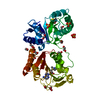
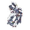
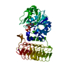
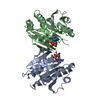
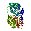
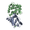
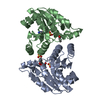
| Title | Crystal structure of C2498 2'-O-ribose methyltransferase RlmM from Escherichia coli | ||||||
 Components Components | RIBOSOMAL RNA LARGE SUBUNIT METHYLTRANSFERASE M | ||||||
 Keywords Keywords | TRANSFERASE | ||||||
| Function / homology |  Function and homology information23S rRNA (cytidine2498-2'-O)-methyltransferase / rRNA (cytosine-2'-O-)-methyltransferase activity / cytoplasm Function and homology information23S rRNA (cytidine2498-2'-O)-methyltransferase / rRNA (cytosine-2'-O-)-methyltransferase activity / cytoplasmSimilarity search - Function | ||||||
| Biological species |  ESCHERICHIA COLI (E. coli) ESCHERICHIA COLI (E. coli) | ||||||
| Method | X-RAY DIFFRACTION / SYNCHROTRON / MOLECULAR REPLACEMENT / Resolution: 1.95 Å | ||||||
 Authors Authors | Punekar, A.S. / Shepherd, T.R. / Liljeruhm, J. / Forster, A.C. / Selmer, M. | ||||||
 Citation Citation | Journal: Nucleic Acids Res. / Year: 2012 Title: Crystal Structure of Rlmm, the 2'O-Ribose Methyltransferase for C2498 of Escherichia Coli 23S Rrna. Authors: Punekar, A.S. / Shepherd, T.R. / Liljeruhm, J. / Forster, A.C. / Selmer, M. | ||||||
| History |
|
- Structure visualization
| Structure viewer | Molecule: MolmilJmol/JSmol |
|---|
- Downloads & links
-Download
| PDBx/mmCIF format | 4atn.cif.gz | 93.9 KB | Display | PDBx/mmCIF format |
|---|---|---|---|---|
| PDB format | pdb4atn.ent.gz | 71.2 KB | Display | PDB format |
| PDBx/mmJSON format | 4atn.json.gz | Tree view | PDBx/mmJSON format | |
| Others |  Other downloads Other downloads |
-Validation report
| Arichive directory | https://data.pdbj.org/pub/pdb/validation_reports/at/4atnftp://data.pdbj.org/pub/pdb/validation_reports/at/4atn | HTTPS FTP |
|---|
-Related structure data


-Links
 PDBj
PDBj

- Assembly
| Deposited unit | 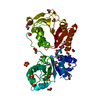
| ||||||||
|---|---|---|---|---|---|---|---|---|---|
| 1 |
| ||||||||
| Unit cell |
|
-Components
| #1: Protein | / 23S RRNA (CYTIDINE2498-2'-O)-METHYLTRANSFERASE / 23S RRNA 2'-O-RIBOSE METHYLTRANSFERASE RLMM Mass: 43092.406 Da / Num. of mol.: 1 Source method: isolated from a genetically manipulated source Source: (gene. exp.) ESCHERICHIA COLI (E. coli) / Strain: K-12 / Plasmid: PEXP5-CT/TOPO / Production host: ESCHERICHIA COLI (E. coli) / Strain (production host): BL21(DE3)References: UniProt: P0ADR6, 23S rRNA (cytidine2498-2'-O)-methyltransferase | ||||||||||
|---|---|---|---|---|---|---|---|---|---|---|---|
| #2: Chemical | ChemComp-GOL / Glycerol  Mass: 92.094 Da / Num. of mol.: 5 / Source method: obtained synthetically / Formula: C3H8O3 Mass: 92.094 Da / Num. of mol.: 5 / Source method: obtained synthetically / Formula: C3H8O3#3: Chemical | ChemComp-EDO / Ethylene glycol  Mass: 62.068 Da / Num. of mol.: 21 / Source method: obtained synthetically / Formula: C2H6O2 Mass: 62.068 Da / Num. of mol.: 21 / Source method: obtained synthetically / Formula: C2H6O2#4: Chemical | ChemComp-SO4 / Sulfate  Mass: 96.063 Da / Num. of mol.: 4 / Source method: obtained synthetically / Formula: SO4 Mass: 96.063 Da / Num. of mol.: 4 / Source method: obtained synthetically / Formula: SO4#5: Water | ChemComp-HOH / | Water Mass: 18.015 Da / Num. of mol.: 195 / Source method: isolated from a natural source / Formula: H2O Mass: 18.015 Da / Num. of mol.: 195 / Source method: isolated from a natural source / Formula: H2OCompound details | CATALYZES THE 2'-O-METHYLATIO | Sequence details | HAS 6XHIS-TAG AT THE C-TERMINAL END | |
-Experimental details
-Experiment
| Experiment | Method: X-RAY DIFFRACTION / Number of used crystals: 1 |
|---|
- Sample preparation
| Crystal | Density Matthews: 2.4 Å3/Da / Density % sol: 48 % / Description: NONE |
|---|---|
| Crystal grow | pH: 8.5 Details: 0.3 M AMMONIUM TARTRATE DIBASIC PH 7.0, 20% PEG 3350, 3.0% DEXTRAN SULFATE SODIUM SALT |
-Data collection
| Diffraction | Mean temperature: 100 K |
|---|---|
| Diffraction source | Source: SYNCHROTRON / Site: MAX II  / Beamline: I911-2 / Wavelength: 1.04 / Beamline: I911-2 / Wavelength: 1.04 |
| Detector | Type: MARRESEARCH SX-165 / Detector: CCD / Date: Mar 7, 2012 |
| Radiation | Monochromator: BENT SI (111) CRYSTAL, HORIZONTALLY FOCUSING OPTICS MULTILAYER MIRROR, CURVED TO FOCUS IN THE VERTICAL Protocol: SINGLE WAVELENGTH / Monochromatic (M) / Laue (L): M / Scattering type: x-ray |
| Radiation wavelength | Wavelength: 1.04 Å / Relative weight: 1 |
| Reflection | Resolution: 1.95→28.77 Å / Num. obs: 30022 / % possible obs: 99.8 % / Observed criterion σ(I): 2 / Redundancy: 7.3 % / Biso Wilson estimate: 33.02 Å2 / Rmerge(I) obs: 0.06 / Net I/σ(I): 25 |
| Reflection shell | Resolution: 1.95→2 Å / Redundancy: 6.3 % / Rmerge(I) obs: 0.87 / Mean I/σ(I) obs: 3.5 / % possible all: 96 |
- Processing
| Software |
| ||||||||||||||||||||||||||||||||||||||||||||||||||||||||||||||||||||||||||||||||||||
|---|---|---|---|---|---|---|---|---|---|---|---|---|---|---|---|---|---|---|---|---|---|---|---|---|---|---|---|---|---|---|---|---|---|---|---|---|---|---|---|---|---|---|---|---|---|---|---|---|---|---|---|---|---|---|---|---|---|---|---|---|---|---|---|---|---|---|---|---|---|---|---|---|---|---|---|---|---|---|---|---|---|---|---|---|---|
| Refinement | Method to determine structure: MOLECULAR REPLACEMENT Starting model: MODEL OBTAINED FROM K2PTCL4 DERIVATIVE CRYSTAL DATA Resolution: 1.95→28.769 Å / SU ML: 0.27 / σ(F): 1.99 / Phase error: 19.82 / Stereochemistry target values: ML
| ||||||||||||||||||||||||||||||||||||||||||||||||||||||||||||||||||||||||||||||||||||
| Solvent computation | Shrinkage radii: 0.86 Å / VDW probe radii: 1.1 Å / Solvent model: FLAT BULK SOLVENT MODEL / Bsol: 69.848 Å2 / ksol: 0.4 e/Å3 | ||||||||||||||||||||||||||||||||||||||||||||||||||||||||||||||||||||||||||||||||||||
| Displacement parameters | Biso mean: 34.12 Å2
| ||||||||||||||||||||||||||||||||||||||||||||||||||||||||||||||||||||||||||||||||||||
| Refinement step | Cycle: LAST / Resolution: 1.95→28.769 Å
| ||||||||||||||||||||||||||||||||||||||||||||||||||||||||||||||||||||||||||||||||||||
| Refine LS restraints |
| ||||||||||||||||||||||||||||||||||||||||||||||||||||||||||||||||||||||||||||||||||||
| LS refinement shell |
|
