 Movie
Movie Controller

[English] 日本語
 Yorodumi
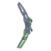
- PDB-4atm: Crystal structure of the BAR domain of human Amphiphysin, isoform... -
+ Open data

- Basic information
| Entry | Database: PDB / ID: 4atm | |||||||||
|---|---|---|---|---|---|---|---|---|---|---|
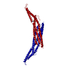
| Title | Crystal structure of the BAR domain of human Amphiphysin, isoform 1 at 1.8 Angstrom resolution featuring increased order at the N- terminus. | |||||||||
 Components Components | AMPHIPHYSIN | |||||||||
 Keywords Keywords | STRUCTURAL PROTEIN / INVAGINATION / KNOBS-IN-HOLES / STRUCTURAL GENOMICS CONSORTIUM / SGC | |||||||||
| Function / homology |  Function and homology information Function and homology informationleading edge membrane / synaptic vesicle endocytosis / phospholipid binding / synaptic vesicle membrane / endocytosis / synaptic vesicle / actin cytoskeleton / Clathrin-mediated endocytosis / chemical synaptic transmission / plasma membrane / cytosolSimilarity search - Function | |||||||||
| Biological species |  HOMO SAPIENS (human) HOMO SAPIENS (human) | |||||||||
| Method | X-RAY DIFFRACTION / SYNCHROTRON / MOLECULAR REPLACEMENT / Resolution: 1.783 Å | |||||||||
 Authors Authors | Allerston, C.K. / Krojer, T. / Arrowsmith, C.H. / Weigelt, J. / Edwards, A. / Bountra, C. / von Delft, F. / Gileadi, O. | |||||||||
 Citation Citation | Journal: To be Published Title: Crystal Structure of the Bar Domain of Human Amphiphysin, Isoform 1 Authors: Allerston, C.K. / Krojer, T. / Gileadi, O. | |||||||||
| History |
|
- Structure visualization
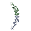
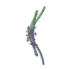
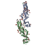
| Structure viewer | Molecule: MolmilJmol/JSmol |
|---|
- Downloads & links
-Download
| PDBx/mmCIF format | 4atm.cif.gz | 113.4 KB | Display | PDBx/mmCIF format |
|---|---|---|---|---|
| PDB format | pdb4atm.ent.gz | 87.7 KB | Display | PDB format |
| PDBx/mmJSON format | 4atm.json.gz | Tree view | PDBx/mmJSON format | |
| Others |  Other downloads Other downloads |
-Validation report
| Arichive directory | https://data.pdbj.org/pub/pdb/validation_reports/at/4atmftp://data.pdbj.org/pub/pdb/validation_reports/at/4atm | HTTPS FTP |
|---|
-Related structure data
| Related structure data | 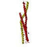 3sogS S: Starting model for refinement |
|---|---|
| Similar structure data |


-Links
 PDBj
PDBj
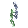
- Assembly
| Deposited unit | 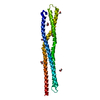
| |||||||||
|---|---|---|---|---|---|---|---|---|---|---|
| 1 | 
| |||||||||
| Unit cell |
| |||||||||
| Components on special symmetry positions |
|
-Components
| #1: Protein | Mass: 28545.377 Da / Num. of mol.: 1 / Fragment: RESIDUES 1-242 Source method: isolated from a genetically manipulated source Source: (gene. exp.) HOMO SAPIENS (human) / Plasmid: PNIC28-BSA4 / Production host:  ESCHERICHIA COLI (E. coli) / Strain (production host): BL21(DE3) / Variant (production host): R3-PRARE2 / References: UniProt: P49418 ESCHERICHIA COLI (E. coli) / Strain (production host): BL21(DE3) / Variant (production host): R3-PRARE2 / References: UniProt: P49418 | ||||||
|---|---|---|---|---|---|---|---|
| #2: Chemical | ChemComp-EDO / Ethylene glycol  Mass: 62.068 Da / Num. of mol.: 5 / Source method: obtained synthetically / Formula: C2H6O2 Mass: 62.068 Da / Num. of mol.: 5 / Source method: obtained synthetically / Formula: C2H6O2#3: Chemical | Glycerol  Mass: 92.094 Da / Num. of mol.: 2 / Source method: obtained synthetically / Formula: C3H8O3 Mass: 92.094 Da / Num. of mol.: 2 / Source method: obtained synthetically / Formula: C3H8O3#4: Water | ChemComp-HOH / | Water Mass: 18.015 Da / Num. of mol.: 233 / Source method: isolated from a natural source / Formula: H2O Mass: 18.015 Da / Num. of mol.: 233 / Source method: isolated from a natural source / Formula: H2OSequence details | G116E MUTATION DERIVED DURING PCR | |
-Experimental details
-Experiment
| Experiment | Method: X-RAY DIFFRACTION / Number of used crystals: 1 |
|---|
- Sample preparation
| Crystal | Density Matthews: 2.99 Å3/Da / Density % sol: 58.82 % / Description: NONE |
|---|---|
| Crystal grow | pH: 7 / Details: 20% PEG 3350, 0.1 M CITRATE PH 5.5 |
-Data collection
| Diffraction | Mean temperature: 100 K |
|---|---|
| Diffraction source | Source: SYNCHROTRON / Site: Diamond  / Beamline: I04 / Wavelength: 0.9795 / Beamline: I04 / Wavelength: 0.9795 |
| Detector | Type: ADSC QUANTUM 315 / Detector: CCD / Date: Jul 21, 2011 / Details: MIRRORS |
| Radiation | Monochromator: DOUBLE CRYSTAL / Protocol: SINGLE WAVELENGTH / Monochromatic (M) / Laue (L): M / Scattering type: x-ray |
| Radiation wavelength | Wavelength: 0.9795 Å / Relative weight: 1 |
| Reflection | Resolution: 1.78→37.2 Å / Num. obs: 32882 / % possible obs: 100 % / Redundancy: 7.6 % / Biso Wilson estimate: 25.89 Å2 / Rmerge(I) obs: 0.09 / Net I/σ(I): 12.8 |
| Reflection shell | Resolution: 1.78→1.88 Å / Redundancy: 7.9 % / Rmerge(I) obs: 0.97 / Mean I/σ(I) obs: 2.2 / % possible all: 100 |
- Processing
| Software |
| ||||||||||||||||||||||||||||||||||||||||||||||||||||||||||||||||||||||||||||||||||||||||||||||||||||||||||||||||||||||||||||||||||||||||||||||||||||||
|---|---|---|---|---|---|---|---|---|---|---|---|---|---|---|---|---|---|---|---|---|---|---|---|---|---|---|---|---|---|---|---|---|---|---|---|---|---|---|---|---|---|---|---|---|---|---|---|---|---|---|---|---|---|---|---|---|---|---|---|---|---|---|---|---|---|---|---|---|---|---|---|---|---|---|---|---|---|---|---|---|---|---|---|---|---|---|---|---|---|---|---|---|---|---|---|---|---|---|---|---|---|---|---|---|---|---|---|---|---|---|---|---|---|---|---|---|---|---|---|---|---|---|---|---|---|---|---|---|---|---|---|---|---|---|---|---|---|---|---|---|---|---|---|---|---|---|---|---|---|---|---|
| Refinement | Method to determine structure: MOLECULAR REPLACEMENT Starting model: PDB ENTRY 3SOG Resolution: 1.783→37.014 Å / SU ML: 0.22 / σ(F): 1.33 / Phase error: 20.48 / Stereochemistry target values: ML
| ||||||||||||||||||||||||||||||||||||||||||||||||||||||||||||||||||||||||||||||||||||||||||||||||||||||||||||||||||||||||||||||||||||||||||||||||||||||
| Solvent computation | Shrinkage radii: 1.11 Å / VDW probe radii: 1.3 Å / Solvent model: FLAT BULK SOLVENT MODEL / Bsol: 36.719 Å2 / ksol: 0.323 e/Å3 | ||||||||||||||||||||||||||||||||||||||||||||||||||||||||||||||||||||||||||||||||||||||||||||||||||||||||||||||||||||||||||||||||||||||||||||||||||||||
| Displacement parameters |
| ||||||||||||||||||||||||||||||||||||||||||||||||||||||||||||||||||||||||||||||||||||||||||||||||||||||||||||||||||||||||||||||||||||||||||||||||||||||
| Refinement step | Cycle: LAST / Resolution: 1.783→37.014 Å
| ||||||||||||||||||||||||||||||||||||||||||||||||||||||||||||||||||||||||||||||||||||||||||||||||||||||||||||||||||||||||||||||||||||||||||||||||||||||
| Refine LS restraints |
| ||||||||||||||||||||||||||||||||||||||||||||||||||||||||||||||||||||||||||||||||||||||||||||||||||||||||||||||||||||||||||||||||||||||||||||||||||||||
| LS refinement shell |
| ||||||||||||||||||||||||||||||||||||||||||||||||||||||||||||||||||||||||||||||||||||||||||||||||||||||||||||||||||||||||||||||||||||||||||||||||||||||
| Refinement TLS params. | Method: refined / Refine-ID: X-RAY DIFFRACTION
| ||||||||||||||||||||||||||||||||||||||||||||||||||||||||||||||||||||||||||||||||||||||||||||||||||||||||||||||||||||||||||||||||||||||||||||||||||||||
| Refinement TLS group |
|
