 Movie
Movie Controller

[English] 日本語
 Yorodumi
- PDB-4ari: Ternary complex of E. coli leucyl-tRNA synthetase, tRNA(leu) and ... -
+ Open data

- Basic information
| Entry | Database: PDB / ID: 4ari | |||||||||
|---|---|---|---|---|---|---|---|---|---|---|
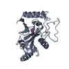
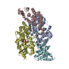
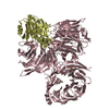
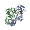
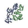
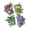
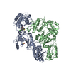
| Title | Ternary complex of E. coli leucyl-tRNA synthetase, tRNA(leu) and the benzoxaborole AN2679 in the editing conformation | |||||||||
 Components Components |
| |||||||||
 Keywords Keywords | LIGASE/RNA / LIGASE-RNA COMPLEX /  LIGASE / NUCLEOTIDE(ATP)-BINDING / PROTEIN BIOSYNTHESIS / CLASS I AMINOACYL-TRNA SYNTHETASE LIGASE / NUCLEOTIDE(ATP)-BINDING / PROTEIN BIOSYNTHESIS / CLASS I AMINOACYL-TRNA SYNTHETASE | |||||||||
| Function / homology |  Function and homology informationleucine-tRNA ligase / leucine-tRNA ligase activity / leucyl-tRNA aminoacylation / aminoacyl-tRNA editing activity / ATP binding / cytosol Function and homology informationleucine-tRNA ligase / leucine-tRNA ligase activity / leucyl-tRNA aminoacylation / aminoacyl-tRNA editing activity / ATP binding / cytosolSimilarity search - Function | |||||||||
| Biological species |  ESCHERICHIA COLI (E. coli) ESCHERICHIA COLI (E. coli) | |||||||||
| Method | X-RAY DIFFRACTION / SYNCHROTRON / MOLECULAR REPLACEMENT / Resolution: 2.08 Å | |||||||||
 Authors Authors | Palencia, A. / Crepin, T. / Vu, M.T. / Lincecum Jr, T.L. / Martinis, S.A. / Cusack, S. | |||||||||
 Citation Citation | Journal: Nat.Struct.Mol.Biol. / Year: 2012 Title: Structural Dynamics of the Aminoacylation and Proofreading Functional Cycle of Bacterial Leucyl-tRNA Synthetase Authors: Palencia, A. / Crepin, T. / Vu, M.T. / Lincecum Jr, T.L. / Martinis, S.A. / Cusack, S. | |||||||||
| History |
|
- Structure visualization
| Structure viewer | Molecule: MolmilJmol/JSmol |
|---|
- Downloads & links
-Download
| PDBx/mmCIF format | 4ari.cif.gz | 232.8 KB | Display | PDBx/mmCIF format |
|---|---|---|---|---|
| PDB format | pdb4ari.ent.gz | 176.3 KB | Display | PDB format |
| PDBx/mmJSON format | 4ari.json.gz | Tree view | PDBx/mmJSON format | |
| Others |  Other downloads Other downloads |
-Validation report
| Arichive directory | https://data.pdbj.org/pub/pdb/validation_reports/ar/4ariftp://data.pdbj.org/pub/pdb/validation_reports/ar/4ari | HTTPS FTP |
|---|
-Related structure data
| Related structure data |  4aq7C  4arcC  4as1C 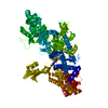 1h3nS C: citing same article ( S: Starting model for refinement |
|---|---|
| Similar structure data |


-Links
 PDBj
PDBj


- Assembly
| Deposited unit | 
| ||||||||
|---|---|---|---|---|---|---|---|---|---|
| 1 |
| ||||||||
| Unit cell |
|
-Components
| #1: Protein | Mass: 99516.016 Da / Num. of mol.: 1 Source method: isolated from a genetically manipulated source Source: (gene. exp.) ESCHERICHIA COLI (E. coli) / Strain: K-12 / Production host: ESCHERICHIA COLI (E. coli) / Strain (production host): BL21 / References: UniProt: P07813, leucine-tRNA ligase | ||||
|---|---|---|---|---|---|
| #2: RNA chain | Mass: 28167.570 Da / Num. of mol.: 1 / Source method: obtained synthetically / Details: UNMODIFIED T7 TRANSCRIPT / Source: (synth.) ESCHERICHIA COLI (E. coli) | ||||
| #3: Chemical | ChemComp-GOL / Glycerol  Mass: 92.094 Da / Num. of mol.: 4 / Source method: obtained synthetically / Formula: C3H8O3 Mass: 92.094 Da / Num. of mol.: 4 / Source method: obtained synthetically / Formula: C3H8O3#4: Chemical |   Mass: 24.305 Da / Num. of mol.: 2 / Source method: obtained synthetically / Formula: Mg Mass: 24.305 Da / Num. of mol.: 2 / Source method: obtained synthetically / Formula: Mg#5: Water | ChemComp-HOH / | Water Mass: 18.015 Da / Num. of mol.: 324 / Source method: isolated from a natural source / Formula: H2O Mass: 18.015 Da / Num. of mol.: 324 / Source method: isolated from a natural source / Formula: H2O |
-Experimental details
-Experiment
| Experiment | Method: X-RAY DIFFRACTION / Number of used crystals: 1 |
|---|
- Sample preparation
| Crystal | Density Matthews: 2.5 Å3/Da / Density % sol: 49.8 % / Description: NONE |
|---|---|
| Crystal grow | pH: 5.6 Details: THE TERNARY COMPLEX ECLEURS-TRNA-AN2679 WAS CRYSTALLIZED FROM 0.1 M SODIUM ACETATE (PH 5.6), 14-18% (W/V) PEG 6000 AND 20 MM NACL. THE CRYSTALS WERE FROZEN IN LIQUID NITROGEN USING 22% (V/V) ...Details: THE TERNARY COMPLEX ECLEURS-TRNA-AN2679 WAS CRYSTALLIZED FROM 0.1 M SODIUM ACETATE (PH 5.6), 14-18% (W/V) PEG 6000 AND 20 MM NACL. THE CRYSTALS WERE FROZEN IN LIQUID NITROGEN USING 22% (V/V) ETHYLENE GLYCOL AS CRYOPROTECTANT |
-Data collection
| Diffraction | Mean temperature: 100 K |
|---|---|
| Diffraction source | Source: SYNCHROTRON / Site: ESRF  / Beamline: ID14-3 / Wavelength: 0.931 / Beamline: ID14-3 / Wavelength: 0.931 |
| Detector | Type: ADSC QUANTUM 4 / Detector: CCD / Date: Aug 27, 2007 |
| Radiation | Protocol: SINGLE WAVELENGTH / Monochromatic (M) / Laue (L): M / Scattering type: x-ray |
| Radiation wavelength | Wavelength: 0.931 Å / Relative weight: 1 |
| Reflection | Resolution: 2.08→50 Å / Num. obs: 74893 / % possible obs: 96.7 % / Observed criterion σ(I): 0 / Redundancy: 3.91 % / Rmerge(I) obs: 0.06 / Net I/σ(I): 20.1 |
| Reflection shell | Resolution: 2.08→2.15 Å / Redundancy: 2.8 % / Rmerge(I) obs: 0.37 / Mean I/σ(I) obs: 3.1 / % possible all: 74.2 |
- Processing
| Software |
| ||||||||||||||||||||||||||||||||||||||||||||||||||||||||||||||||||||||||||||||||||||||||||||||||||||||||||||||||||||||||||||||||||||||||||||||||||||||||||||||||||||||||||||||||||||||
|---|---|---|---|---|---|---|---|---|---|---|---|---|---|---|---|---|---|---|---|---|---|---|---|---|---|---|---|---|---|---|---|---|---|---|---|---|---|---|---|---|---|---|---|---|---|---|---|---|---|---|---|---|---|---|---|---|---|---|---|---|---|---|---|---|---|---|---|---|---|---|---|---|---|---|---|---|---|---|---|---|---|---|---|---|---|---|---|---|---|---|---|---|---|---|---|---|---|---|---|---|---|---|---|---|---|---|---|---|---|---|---|---|---|---|---|---|---|---|---|---|---|---|---|---|---|---|---|---|---|---|---|---|---|---|---|---|---|---|---|---|---|---|---|---|---|---|---|---|---|---|---|---|---|---|---|---|---|---|---|---|---|---|---|---|---|---|---|---|---|---|---|---|---|---|---|---|---|---|---|---|---|---|---|
| Refinement | Method to determine structure: MOLECULAR REPLACEMENT Starting model: PDB ENTRY 1H3N Resolution: 2.08→90.91 Å / Cor.coef. Fo:Fc: 0.941 / Cor.coef. Fo:Fc free: 0.912 / SU B: 4.66 / SU ML: 0.125 / Cross valid method: THROUGHOUT / ESU R: 0.206 / ESU R Free: 0.182 / Stereochemistry target values: MAXIMUM LIKELIHOOD Details: HYDROGENS HAVE BEEN ADDED IN THE RIDING POSITIONS. U VALUES REFINED INDIVIDUALLY
| ||||||||||||||||||||||||||||||||||||||||||||||||||||||||||||||||||||||||||||||||||||||||||||||||||||||||||||||||||||||||||||||||||||||||||||||||||||||||||||||||||||||||||||||||||||||
| Solvent computation | Ion probe radii: 0.8 Å / Shrinkage radii: 0.8 Å / VDW probe radii: 1.2 Å / Solvent model: MASK | ||||||||||||||||||||||||||||||||||||||||||||||||||||||||||||||||||||||||||||||||||||||||||||||||||||||||||||||||||||||||||||||||||||||||||||||||||||||||||||||||||||||||||||||||||||||
| Displacement parameters | Biso mean: 30.479 Å2
| ||||||||||||||||||||||||||||||||||||||||||||||||||||||||||||||||||||||||||||||||||||||||||||||||||||||||||||||||||||||||||||||||||||||||||||||||||||||||||||||||||||||||||||||||||||||
| Refinement step | Cycle: LAST / Resolution: 2.08→90.91 Å
| ||||||||||||||||||||||||||||||||||||||||||||||||||||||||||||||||||||||||||||||||||||||||||||||||||||||||||||||||||||||||||||||||||||||||||||||||||||||||||||||||||||||||||||||||||||||
| Refine LS restraints |
|