 Movie
Movie Controller


+ Open data

- Basic information
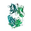
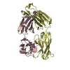
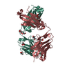
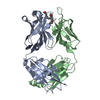
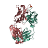
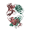
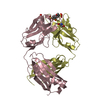
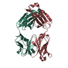
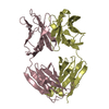
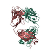
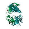
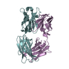
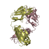
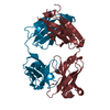
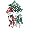
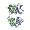
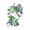
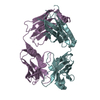
| Entry | Database: PDB / ID: 4amk | ||||||
|---|---|---|---|---|---|---|---|
| Title | Fab fragment of antiporphrin antibody 13g10 | ||||||
 Components Components |
| ||||||
 Keywords Keywords |  IMMUNE SYSTEM / 13G10 / METALLOPORPHYRIN / CATALYTIC ANTIBODY / PEROXIDASE IMMUNE SYSTEM / 13G10 / METALLOPORPHYRIN / CATALYTIC ANTIBODY / PEROXIDASE | ||||||
| Function / homology | Immunoglobulins / Immunoglobulin-like / Sandwich / Mainly Beta Function and homology information Function and homology information | ||||||
| Biological species |  MUS MUSCULUS (house mouse) MUS MUSCULUS (house mouse) | ||||||
| Method | X-RAY DIFFRACTION / SYNCHROTRON / MOLECULAR REPLACEMENT / Resolution: 2.05 Å | ||||||
 Authors Authors | Mahy, J.P. / Golinelli-Pimpaneau, B. | ||||||
 Citation Citation | Journal: Plos One / Year: 2012 Title: Crystal Structure of Two Anti-Porphyrin Antibodies with Peroxidase Activity. Authors: Munoz Robles, V. / Marechal, J. / Bahloul, A. / Sari, M. / Mahy, J. / Golinelli-Pimpaneau, B. #1: Journal: FEBS Lett. / Year: 1996 Title: Artificial Peroxidase-Like Hemoproteins Based on Antibodies Constructed from a Specifically Designed Ortho-Carboxy Substituted Tetraarylporphyrin Hapten and Exhibiting a High Affinity for Iron-Porphyrins. Authors: Quilez, R. / De Lauzon, S. / Desfosses, B. / Mansuy, D. / Mahy, J.P. #2: Journal: FEBS Lett. / Year: 1999 Title: Studies of the Reactivity of Artificial Peroxidase-Like Hemoproteins Based on Antibodies Elicited Against a Specifically Designed Ortho-Carboxy Substituted Tetraarylporphyrin. Authors: De Lauzon, S. / Desfosses, B. / Mansuy, D. / Mahy, J.P. | ||||||
| History |
|
- Structure visualization
| Structure viewer | Molecule: MolmilJmol/JSmol |
|---|
- Downloads & links
-Download
| PDBx/mmCIF format | 4amk.cif.gz | 99.4 KB | Display | PDBx/mmCIF format |
|---|---|---|---|---|
| PDB format | pdb4amk.ent.gz | 74.5 KB | Display | PDB format |
| PDBx/mmJSON format | 4amk.json.gz | Tree view | PDBx/mmJSON format | |
| Others |  Other downloads Other downloads |
-Validation report
| Arichive directory | https://data.pdbj.org/pub/pdb/validation_reports/am/4amkftp://data.pdbj.org/pub/pdb/validation_reports/am/4amk | HTTPS FTP |
|---|
-Related structure data
| Related structure data |  4at6C 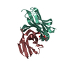 1mfaS 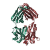 1mfeS S: Starting model for refinement C: citing same article ( |
|---|---|
| Similar structure data |

-Links
 PDBj
PDBj

- Assembly
| Deposited unit | 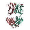
| ||||||||
|---|---|---|---|---|---|---|---|---|---|
| 1 |
| ||||||||
| Unit cell |
|
-Components
| #1: Antibody | Mass: 22877.721 Da / Num. of mol.: 1 / Source method: isolated from a natural source / Source: (natural) MUS MUSCULUS (house mouse) / Cell line: MURINE HYBRIDOMA | ||||
|---|---|---|---|---|---|
| #2: Antibody | Mass: 22751.240 Da / Num. of mol.: 1 / Source method: isolated from a natural source / Source: (natural) MUS MUSCULUS (house mouse) / Cell line: MURINE HYBRIDOMA | ||||
| #3: Chemical | Glycerol  Mass: 92.094 Da / Num. of mol.: 3 / Source method: obtained synthetically / Formula: C3H8O3 Mass: 92.094 Da / Num. of mol.: 3 / Source method: obtained synthetically / Formula: C3H8O3#4: Chemical | ChemComp-MG / |   Mass: 24.305 Da / Num. of mol.: 1 / Source method: obtained synthetically / Formula: Mg Mass: 24.305 Da / Num. of mol.: 1 / Source method: obtained synthetically / Formula: Mg#5: Water | ChemComp-HOH / | Water Mass: 18.015 Da / Num. of mol.: 188 / Source method: isolated from a natural source / Formula: H2O Mass: 18.015 Da / Num. of mol.: 188 / Source method: isolated from a natural source / Formula: H2O |
-Experimental details
-Experiment
| Experiment | Method: X-RAY DIFFRACTION / Number of used crystals: 1 |
|---|
- Sample preparation
| Crystal | Density Matthews: 1.99 Å3/Da / Density % sol: 38 % / Description: NONE |
|---|---|
| Crystal grow | pH: 8.5 Details: 26.5 % PEG 2000, 0.2 M MGCL2, 0.1 M TRIS PH 8.5, 10% GLYCEROL |
-Data collection
| Diffraction | Mean temperature: 100 K |
|---|---|
| Diffraction source | Source: SYNCHROTRON / Site: ESRF  / Beamline: ID14-1 / Wavelength: 0.934 / Beamline: ID14-1 / Wavelength: 0.934 |
| Detector | Type: ADSC QUANTUM 210 / Detector: CCD / Date: Feb 8, 2001 |
| Radiation | Monochromator: 0.97 / Protocol: SINGLE WAVELENGTH / Monochromatic (M) / Laue (L): M / Scattering type: x-ray |
| Radiation wavelength | Wavelength: 0.934 Å / Relative weight: 1 |
| Reflection | Resolution: 2.05→25 Å / Num. obs: 23580 / % possible obs: 93.7 % / Redundancy: 2.5 % / Biso Wilson estimate: 42.5 Å2 / Rmerge(I) obs: 0.04 / Net I/σ(I): 23.7 |
| Reflection shell | Resolution: 2.05→2.09 Å / Redundancy: 2.1 % / Rmerge(I) obs: 0.19 / Mean I/σ(I) obs: 4.6 / % possible all: 91.4 |
- Processing
| Software |
| ||||||||||||||||||||||||||||||||||||||||||||||||||||||||||||
|---|---|---|---|---|---|---|---|---|---|---|---|---|---|---|---|---|---|---|---|---|---|---|---|---|---|---|---|---|---|---|---|---|---|---|---|---|---|---|---|---|---|---|---|---|---|---|---|---|---|---|---|---|---|---|---|---|---|---|---|---|---|
| Refinement | Method to determine structure: MOLECULAR REPLACEMENT Starting model: PDB ENTRIES 1MFA AND 1MFE Resolution: 2.05→20 Å / Data cutoff high absF: 10000 / Cross valid method: THROUGHOUT / σ(F): 0 / Stereochemistry target values: MAXIMUM LIKELIHOOD Details: THE SIDE CHAINS OF SER L32, SER L95 AND GLN H196, WHICH WERE NOT OBSERVED IN THE ELECTRON DENSITY, WERE MODELLED AS ALANINES.
| ||||||||||||||||||||||||||||||||||||||||||||||||||||||||||||
| Solvent computation | Bsol: 69.7339 Å2 / ksol: 0.397451 e/Å3 | ||||||||||||||||||||||||||||||||||||||||||||||||||||||||||||
| Displacement parameters | Biso mean: 49 Å2
| ||||||||||||||||||||||||||||||||||||||||||||||||||||||||||||
| Refinement step | Cycle: LAST / Resolution: 2.05→20 Å
| ||||||||||||||||||||||||||||||||||||||||||||||||||||||||||||
| Refine LS restraints |
| ||||||||||||||||||||||||||||||||||||||||||||||||||||||||||||
| LS refinement shell | Resolution: 2.05→2.08 Å / Total num. of bins used: 22 /
| ||||||||||||||||||||||||||||||||||||||||||||||||||||||||||||
| Xplor file | Serial no: 1 / Param file: GOL.PAR / Topol file: GOL.TOP |
