 Movie
Movie Controller


+ Open data

- Basic information
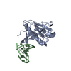
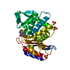
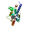
| Entry | Database: PDB / ID: 4al7 | ||||||
|---|---|---|---|---|---|---|---|
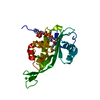
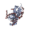
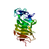
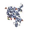
| Title | Crystal structure of the Csy4-minimal crRNA complex | ||||||
 Components Components |
| ||||||
 Keywords Keywords | HYDROLASE/RNA / HYDROLASE-RNA COMPLEX /  CRISPR CRISPR | ||||||
| Function / homology |  Function and homology information Function and homology informationmaintenance of CRISPR repeat elements / endonuclease activity / defense response to virus / Hydrolases; Acting on ester bonds / RNA bindingSimilarity search - Function | ||||||
| Biological species |   PSEUDOMONAS AERUGINOSA (bacteria) PSEUDOMONAS AERUGINOSA (bacteria) | ||||||
| Method | X-RAY DIFFRACTION / SYNCHROTRON / MOLECULAR REPLACEMENT / Resolution: 2.32 Å | ||||||
 Authors Authors | Haurwitz, R.E. / Sternberg, S.H. / Doudna, J.A. | ||||||
 Citation Citation | Journal: Embo J. / Year: 2012 Title: Csy4 Relies on an Unusual Catalytic Dyad to Position and Cleave Crispr RNA. Authors: Haurwitz, R.E. / Sternberg, S.H. / Doudna, J.A. | ||||||
| History |
|
- Structure visualization
| Structure viewer | Molecule: MolmilJmol/JSmol |
|---|
- Downloads & links
-Download
| PDBx/mmCIF format | 4al7.cif.gz | 122.2 KB | Display | PDBx/mmCIF format |
|---|---|---|---|---|
| PDB format | pdb4al7.ent.gz | 93.4 KB | Display | PDB format |
| PDBx/mmJSON format | 4al7.json.gz | Tree view | PDBx/mmJSON format | |
| Others |  Other downloads Other downloads |
-Validation report
| Arichive directory | https://data.pdbj.org/pub/pdb/validation_reports/al/4al7ftp://data.pdbj.org/pub/pdb/validation_reports/al/4al7 | HTTPS FTP |
|---|
-Related structure data
| Related structure data | 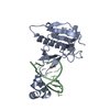 4al5SC  4al6C S: Starting model for refinement C: citing same article ( |
|---|---|
| Similar structure data |


-Links
 PDBj
PDBj


- Assembly
| Deposited unit | 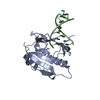
| |||||||||
|---|---|---|---|---|---|---|---|---|---|---|
| 1 |
| |||||||||
| Unit cell |
| |||||||||
| Components on special symmetry positions |
|
-Components
| #1: Protein | Mass: 21819.910 Da / Num. of mol.: 1 Source method: isolated from a genetically manipulated source Source: (gene. exp.) PSEUDOMONAS AERUGINOSA (bacteria) / Strain: UCBPP-PA14 / Production host: ESCHERICHIA COLI (E. coli) / Strain (production host): BL21(DE3) / Variant (production host): ROSETTA 2 / References: UniProt: Q02MM2 |
|---|---|
| #2: RNA chain | Mass: 6359.815 Da / Num. of mol.: 1 / Source method: obtained synthetically / Source: (synth.) PSEUDOMONAS AERUGINOSA (bacteria) |
| #3: Water | ChemComp-HOH / Water Mass: 18.015 Da / Num. of mol.: 45 / Source method: isolated from a natural source / Formula: H2O Mass: 18.015 Da / Num. of mol.: 45 / Source method: isolated from a natural source / Formula: H2O |
| Sequence details | THE UNIPROT DATABASE HAS AN INCORRECT START CODON ANNOTATION. THEREFORE, THE UNIPROT PROTEIN ...THE UNIPROT DATABASE HAS AN INCORRECT START CODON ANNOTATION |
-Experimental details
-Experiment
| Experiment | Method: X-RAY DIFFRACTION / Number of used crystals: 1 |
|---|
- Sample preparation
| Crystal | Density Matthews: 2.37 Å3/Da / Density % sol: 52.24 % / Description: NONE |
|---|---|
| Crystal grow | pH: 5 Details: 21% PEG4000, 180 MM SODIUM CITRATE PH 5.0, 100 MM MAGNESIUM CHLORIDE, 2 MM AMMONIUM METAVANADATE |
-Data collection
| Diffraction | Mean temperature: 100 K |
|---|---|
| Diffraction source | Source: SYNCHROTRON / Site: ALS  / Beamline: 8.2.1 / Wavelength: 0.999982 / Beamline: 8.2.1 / Wavelength: 0.999982 |
| Detector | Type: ADSC QUANTUM 315r / Detector: CCD / Date: May 29, 2011 |
| Radiation | Protocol: SINGLE WAVELENGTH / Monochromatic (M) / Laue (L): M / Scattering type: x-ray |
| Radiation wavelength | Wavelength: 0.999982 Å / Relative weight: 1 |
| Reflection | Resolution: 2.32→36.48 Å / Num. obs: 10773 / % possible obs: 99.4 % / Observed criterion σ(I): 1.99 / Redundancy: 4.9 % / Biso Wilson estimate: 38.68 Å2 / Rmerge(I) obs: 0.08 / Net I/σ(I): 13.5 |
| Reflection shell | Resolution: 2.32→2.38 Å / Redundancy: 3.3 % / Rmerge(I) obs: 0.48 / Mean I/σ(I) obs: 2.49 / % possible all: 100 |
- Processing
| Software |
| |||||||||||||||||||||||||||||||||||||||||||||||||||||||||||||||||||||||||||
|---|---|---|---|---|---|---|---|---|---|---|---|---|---|---|---|---|---|---|---|---|---|---|---|---|---|---|---|---|---|---|---|---|---|---|---|---|---|---|---|---|---|---|---|---|---|---|---|---|---|---|---|---|---|---|---|---|---|---|---|---|---|---|---|---|---|---|---|---|---|---|---|---|---|---|---|---|
| Refinement | Method to determine structure: MOLECULAR REPLACEMENT Starting model: PDB ENTRY 4AL5 Resolution: 2.32→36.476 Å / SU ML: 0.34 / σ(F): 1.99 / Phase error: 27.29 / Stereochemistry target values: ML
| |||||||||||||||||||||||||||||||||||||||||||||||||||||||||||||||||||||||||||
| Solvent computation | Shrinkage radii: 0.86 Å / VDW probe radii: 1.1 Å / Solvent model: FLAT BULK SOLVENT MODEL / Bsol: 59.338 Å2 / ksol: 0.393 e/Å3 | |||||||||||||||||||||||||||||||||||||||||||||||||||||||||||||||||||||||||||
| Displacement parameters | Biso mean: 65.5 Å2
| |||||||||||||||||||||||||||||||||||||||||||||||||||||||||||||||||||||||||||
| Refinement step | Cycle: LAST / Resolution: 2.32→36.476 Å
| |||||||||||||||||||||||||||||||||||||||||||||||||||||||||||||||||||||||||||
| Refine LS restraints |
| |||||||||||||||||||||||||||||||||||||||||||||||||||||||||||||||||||||||||||
| LS refinement shell |
| |||||||||||||||||||||||||||||||||||||||||||||||||||||||||||||||||||||||||||
| Refinement TLS params. | Method: refined / Refine-ID: X-RAY DIFFRACTION
| |||||||||||||||||||||||||||||||||||||||||||||||||||||||||||||||||||||||||||
| Refinement TLS group |
|
