 Movie
Movie Controller

[English] 日本語
 Yorodumi
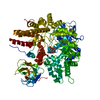
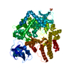
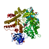
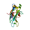
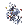
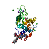
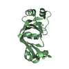
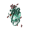
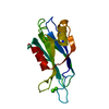
- PDB-4a6o: CpGH89CBM32-4, produced by Clostridium perfringens, in complex wi... -
+ Open data

- Basic information
| Entry | Database: PDB / ID: 4a6o | |||||||||
|---|---|---|---|---|---|---|---|---|---|---|
| Title | CpGH89CBM32-4, produced by Clostridium perfringens, in complex with glcNAc-alpha-1,4-galactose | |||||||||
 Components Components | ALPHA-N-ACETYLGLUCOSAMINIDASE FAMILY PROTEIN | |||||||||
 Keywords Keywords |  HYDROLASE / FAMILY 89 GLYCOSIDE HYDROLASE / FAMILY 32 CARBOHYDRATE-BINDING MODULE CPF_0859 HYDROLASE / FAMILY 89 GLYCOSIDE HYDROLASE / FAMILY 32 CARBOHYDRATE-BINDING MODULE CPF_0859 | |||||||||
| Function / homology |  Function and homology information Function and homology informationhydrolase activity, acting on glycosyl bonds / hydrolase activity, hydrolyzing O-glycosyl compounds / carbohydrate metabolic process / metal ion bindingSimilarity search - Function | |||||||||
| Biological species |   CLOSTRIDIUM PERFRINGENS (bacteria) CLOSTRIDIUM PERFRINGENS (bacteria) | |||||||||
| Method | X-RAY DIFFRACTION / OTHER / Resolution: 2.8 Å | |||||||||
 Authors Authors | Ficko-Blean, E. / Stuart, C.P. / Suits, M.D. / Cid, M. / Tessier, M. / Woods, R.J. / Boraston, A.B. | |||||||||
 Citation Citation | Journal: Plos One / Year: 2012 Title: Carbohydrate Recognition by an Architecturally Complex Alpha-N-Acetylglucosaminidase from Clostridium Perfringens. Authors: Ficko-Blean, E. / Stuart, C.P. / Suits, M.D. / Cid, M. / Tessier, M. / Woods, R.J. / Boraston, A.B. | |||||||||
| History |
|
- Structure visualization
| Structure viewer | Molecule: MolmilJmol/JSmol |
|---|
- Downloads & links
-Download
| PDBx/mmCIF format | 4a6o.cif.gz | 128.2 KB | Display | PDBx/mmCIF format |
|---|---|---|---|---|
| PDB format | pdb4a6o.ent.gz | 100.4 KB | Display | PDB format |
| PDBx/mmJSON format | 4a6o.json.gz | Tree view | PDBx/mmJSON format | |
| Others |  Other downloads Other downloads |
-Validation report
| Arichive directory | https://data.pdbj.org/pub/pdb/validation_reports/a6/4a6oftp://data.pdbj.org/pub/pdb/validation_reports/a6/4a6o | HTTPS FTP |
|---|
-Related structure data
| Related structure data |  4a3zC  4a41C  4a42C  4a44C  4a45C  4aaxC  4a43 C: citing same article ( |
|---|---|
| Similar structure data |


-Links
 PDBj
PDBj


- Assembly
| Deposited unit | 
| |||||||||
|---|---|---|---|---|---|---|---|---|---|---|
| 1 | 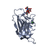
| |||||||||
| 2 | 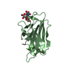
| |||||||||
| Unit cell |
| |||||||||
| Components on special symmetry positions |
|
-Components
| #1: Protein | Mass: 17521.205 Da / Num. of mol.: 2 / Fragment: CBM32-4, RESIDUES 1206-1343 Source method: isolated from a genetically manipulated source Source: (gene. exp.) CLOSTRIDIUM PERFRINGENS (bacteria) / Production host: ESCHERICHIA COLI (E. coli) / Strain (production host): BL21(DE3) / Variant (production host): STARReferences: UniProt: Q0TST1, UniProt: A0A0H2YU91*PLUS, alpha-N-acetylglucosaminidase#2: Polysaccharide | / Mass: 383.349 Da / Num. of mol.: 2Source method: isolated from a genetically manipulated source #3: Chemical |   Mass: 40.078 Da / Num. of mol.: 3 / Source method: obtained synthetically / Formula: Ca Mass: 40.078 Da / Num. of mol.: 3 / Source method: obtained synthetically / Formula: Ca#4: Water | ChemComp-HOH / | Water Mass: 18.015 Da / Num. of mol.: 111 / Source method: isolated from a natural source / Formula: H2O Mass: 18.015 Da / Num. of mol.: 111 / Source method: isolated from a natural source / Formula: H2O |
|---|
-Experimental details
-Experiment
| Experiment | Method: X-RAY DIFFRACTION |
|---|
- Sample preparation
| Crystal | Density Matthews: 2.03 Å3/Da / Density % sol: 39.4 % / Description: NONE |
|---|---|
| Crystal grow | Details: CBM32-4 (20 MG/ML) IN COMPLEX WITH GLCNAC-ALPHA-1,4-GAL (AT 2 MM) CRYSTALLIZED IN 0.1 M ZNOAC, 0.1 M BICINE PH 8.0, 18% PEG 3350, 4 MM CRCL |
-Data collection
| Diffraction | Mean temperature: 113 K |
|---|---|
| Diffraction source | Source: ROTATING ANODE / Wavelength: 1.5418 |
| Radiation | Protocol: SINGLE WAVELENGTH / Monochromatic (M) / Laue (L): M / Scattering type: x-ray |
| Radiation wavelength | Wavelength: 1.5418 Å / Relative weight: 1 |
| Reflection | Resolution: 2.8→20 Å / Num. obs: 7240 / % possible obs: 98.7 % / Observed criterion σ(I): 2 / Redundancy: 4.2 % / Rmerge(I) obs: 0.15 |
- Processing
| Software | Name: REFMAC / Version: 5.5.0109 / Classification: refinement | ||||||||||||||||||||||||||||||||||||||||||||||||||||||||||||||||||||||||||||||||||||||||||||||||||||||||||||||||||||||||||||||||||||||||||||||||||||||||||||||||||||||||||||||||||||||
|---|---|---|---|---|---|---|---|---|---|---|---|---|---|---|---|---|---|---|---|---|---|---|---|---|---|---|---|---|---|---|---|---|---|---|---|---|---|---|---|---|---|---|---|---|---|---|---|---|---|---|---|---|---|---|---|---|---|---|---|---|---|---|---|---|---|---|---|---|---|---|---|---|---|---|---|---|---|---|---|---|---|---|---|---|---|---|---|---|---|---|---|---|---|---|---|---|---|---|---|---|---|---|---|---|---|---|---|---|---|---|---|---|---|---|---|---|---|---|---|---|---|---|---|---|---|---|---|---|---|---|---|---|---|---|---|---|---|---|---|---|---|---|---|---|---|---|---|---|---|---|---|---|---|---|---|---|---|---|---|---|---|---|---|---|---|---|---|---|---|---|---|---|---|---|---|---|---|---|---|---|---|---|---|
| Refinement | Method to determine structure: OTHER Starting model: NONE Resolution: 2.8→20 Å / Cor.coef. Fo:Fc: 0.861 / Cor.coef. Fo:Fc free: 0.854 / SU B: 42.36 / SU ML: 0.388 / Cross valid method: THROUGHOUT / ESU R Free: 0.536 / Stereochemistry target values: MAXIMUM LIKELIHOOD / Details: HYDROGENS HAVE BEEN ADDED IN THE RIDING POSITIONS.
| ||||||||||||||||||||||||||||||||||||||||||||||||||||||||||||||||||||||||||||||||||||||||||||||||||||||||||||||||||||||||||||||||||||||||||||||||||||||||||||||||||||||||||||||||||||||
| Solvent computation | Ion probe radii: 0.8 Å / Shrinkage radii: 0.8 Å / VDW probe radii: 1.4 Å / Solvent model: MASK | ||||||||||||||||||||||||||||||||||||||||||||||||||||||||||||||||||||||||||||||||||||||||||||||||||||||||||||||||||||||||||||||||||||||||||||||||||||||||||||||||||||||||||||||||||||||
| Displacement parameters | Biso mean: 22.208 Å2
| ||||||||||||||||||||||||||||||||||||||||||||||||||||||||||||||||||||||||||||||||||||||||||||||||||||||||||||||||||||||||||||||||||||||||||||||||||||||||||||||||||||||||||||||||||||||
| Refinement step | Cycle: LAST / Resolution: 2.8→20 Å
| ||||||||||||||||||||||||||||||||||||||||||||||||||||||||||||||||||||||||||||||||||||||||||||||||||||||||||||||||||||||||||||||||||||||||||||||||||||||||||||||||||||||||||||||||||||||
| Refine LS restraints |
|