 Movie
Movie Controller


+ Open data

- Basic information
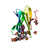
| Entry | Database: PDB / ID: 4a5x | ||||||
|---|---|---|---|---|---|---|---|
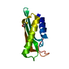
| Title | Structures of MITD1 | ||||||
 Components Components |
| ||||||
 Keywords Keywords |  PROTEIN TRANSPORT / ESCRT / CYTOKINESIS / MIDBODY PROTEIN TRANSPORT / ESCRT / CYTOKINESIS / MIDBODY | ||||||
| Function / homology |  Function and homology information Function and homology informationamphisome membrane / multivesicular body-lysosome fusion / vesicle fusion with vacuole / ESCRT III complex disassembly / late endosome to lysosome transport / ESCRT III complex / kinetochore microtubule / endosome transport via multivesicular body sorting pathway / regulation of centrosome duplication / nuclear membrane reassembly ...amphisome membrane / multivesicular body-lysosome fusion / vesicle fusion with vacuole / ESCRT III complex disassembly / late endosome to lysosome transport / ESCRT III complex / kinetochore microtubule / endosome transport via multivesicular body sorting pathway / regulation of centrosome duplication / nuclear membrane reassembly / midbody abscission / multivesicular body sorting pathway / membrane fission / plasma membrane repair / multivesicular body membrane / late endosome to vacuole transport / ubiquitin-dependent protein catabolic process via the multivesicular body sorting pathway / multivesicular body assembly / regulation of mitotic spindle assembly / mitotic chromosome condensation / microtubule organizing center / nucleus organization / mitotic metaphase chromosome alignment / viral budding via host ESCRT complex / autophagosome maturation / autophagosome membrane / mitotic cytokinesis / endomembrane system / nuclear pore / vesicle-mediated transport / multivesicular body / phosphatidylinositol binding / viral budding from plasma membrane / HCMV Late Events / negative regulation of protein binding / condensed nuclear chromosome / kinetochore / autophagy / nuclear matrix / metallopeptidase activity / protein transport / late endosome membrane / midbody / early endosome / lysosomal membrane / protein domain specific binding / cell division / negative regulation of gene expression / intracellular membrane-bounded organelle / protein homodimerization activity / extracellular exosome / zinc ion binding / membrane / identical protein binding / plasma membrane / cytosolSimilarity search - Function | ||||||
| Biological species |  HOMO SAPIENS (human) HOMO SAPIENS (human) | ||||||
| Method | X-RAY DIFFRACTION / SYNCHROTRON / MOLECULAR REPLACEMENT / Resolution: 1.91 Å | ||||||
 Authors Authors | Hadders, M.A. / Agromayor, M. / Caballe, A. / Obita, T. / Perisic, O. / Williams, R.L. / Martin-Serrano, J. | ||||||
 Citation Citation | Journal: Proc.Natl.Acad.Sci.USA / Year: 2012 Title: Escrt-III Binding Protein Mitd1 is Involved in Cytokinesis and Has an Unanticipated Pld Fold that Binds Membranes. Authors: Hadders, M.A. / Agromayor, M. / Obita, T. / Perisic, O. / Caballe, A. / Kloc, M. / Lamers, M.H. / Williams, R.L. / Martin-Serrano, J. | ||||||
| History |
|
- Structure visualization
| Structure viewer | Molecule: MolmilJmol/JSmol |
|---|
- Downloads & links
-Download
| PDBx/mmCIF format | 4a5x.cif.gz | 87.5 KB | Display | PDBx/mmCIF format |
|---|---|---|---|---|
| PDB format | pdb4a5x.ent.gz | 67.6 KB | Display | PDB format |
| PDBx/mmJSON format | 4a5x.json.gz | Tree view | PDBx/mmJSON format | |
| Others |  Other downloads Other downloads |
-Validation report
| Arichive directory | https://data.pdbj.org/pub/pdb/validation_reports/a5/4a5xftp://data.pdbj.org/pub/pdb/validation_reports/a5/4a5x | HTTPS FTP |
|---|
-Related structure data
| Related structure data | 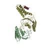 2ymbC  4a5zC  1wfdS S: Starting model for refinement C: citing same article ( |
|---|---|
| Similar structure data |


-Links
 PDBj
PDBj

- Assembly
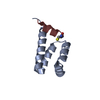
| Deposited unit | 
| ||||||||||||
|---|---|---|---|---|---|---|---|---|---|---|---|---|---|
| 1 | 
| ||||||||||||
| 2 | 
| ||||||||||||
| Unit cell |
| ||||||||||||
| Noncrystallographic symmetry (NCS) | NCS oper:
|
-Components
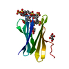
| #1: Protein | Mass: 10053.435 Da / Num. of mol.: 2 / Fragment: MIT, RESIDUES 9-85 Source method: isolated from a genetically manipulated source Source: (gene. exp.) HOMO SAPIENS (human) / Production host:  ESCHERICHIA COLI (E. coli) / Strain (production host): BL21 / Variant (production host): C41 / References: UniProt: Q8WV92 ESCHERICHIA COLI (E. coli) / Strain (production host): BL21 / Variant (production host): C41 / References: UniProt: Q8WV92#2: Protein/peptide | Mass: 1901.157 Da / Num. of mol.: 2 / Fragment: RESIDUES 184-196 Source method: isolated from a genetically manipulated source Source: (gene. exp.) HOMO SAPIENS (human) / Production host: ESCHERICHIA COLI (E. coli) / Strain (production host): BL21 / Variant (production host): C41 / References: UniProt: Q9HD42#3: Chemical | Glycerol  Mass: 92.094 Da / Num. of mol.: 2 / Source method: obtained synthetically / Formula: C3H8O3 Mass: 92.094 Da / Num. of mol.: 2 / Source method: obtained synthetically / Formula: C3H8O3#4: Chemical | ChemComp-P15 / | 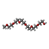  Mass: 296.357 Da / Num. of mol.: 1 / Source method: obtained synthetically / Formula: C13H28O7 Mass: 296.357 Da / Num. of mol.: 1 / Source method: obtained synthetically / Formula: C13H28O7#5: Water | ChemComp-HOH / | Water Mass: 18.015 Da / Num. of mol.: 75 / Source method: isolated from a natural source / Formula: H2O Mass: 18.015 Da / Num. of mol.: 75 / Source method: isolated from a natural source / Formula: H2O |
|---|
-Experimental details
-Experiment
| Experiment | Method: X-RAY DIFFRACTION / Number of used crystals: 1 |
|---|
- Sample preparation
| Crystal | Density Matthews: 2 Å3/Da / Density % sol: 38 % / Description: NONE |
|---|---|
| Crystal grow | Details: 29% PEG2000MME, 160 MM KSCN |
-Data collection
| Diffraction | Mean temperature: 100 K |
|---|---|
| Diffraction source | Source: SYNCHROTRON / Site: Diamond  / Beamline: I03 / Wavelength: 0.9763 / Beamline: I03 / Wavelength: 0.9763 |
| Detector | Type: ADSC CCD / Detector: CCD / Date: Sep 29, 2010 |
| Radiation | Protocol: SINGLE WAVELENGTH / Monochromatic (M) / Laue (L): M / Scattering type: x-ray |
| Radiation wavelength | Wavelength: 0.9763 Å / Relative weight: 1 |
| Reflection | Resolution: 1.91→30 Å / Num. obs: 17144 / % possible obs: 97.6 % / Observed criterion σ(I): 2 / Redundancy: 7.3 % / Biso Wilson estimate: 31.13 Å2 / Rmerge(I) obs: 0.06 / Net I/σ(I): 22.5 |
| Reflection shell | Resolution: 1.91→2.02 Å / Redundancy: 7.3 % / Rmerge(I) obs: 0.62 / Mean I/σ(I) obs: 3.2 / % possible all: 94.6 |
- Processing
| Software |
| |||||||||||||||||||||||||||||||||||||||||||||||||||||||||||||||||||||||||||||||||||||||||||||||||||||||||||||||||||||||||||||||||||||||||||||||||||||||||||||||||||||||||||||||||||||||||||||||||||||||||||||||||||||||||||||||||||||||||||||||||||||||||||||||||||||||||||||||||||
|---|---|---|---|---|---|---|---|---|---|---|---|---|---|---|---|---|---|---|---|---|---|---|---|---|---|---|---|---|---|---|---|---|---|---|---|---|---|---|---|---|---|---|---|---|---|---|---|---|---|---|---|---|---|---|---|---|---|---|---|---|---|---|---|---|---|---|---|---|---|---|---|---|---|---|---|---|---|---|---|---|---|---|---|---|---|---|---|---|---|---|---|---|---|---|---|---|---|---|---|---|---|---|---|---|---|---|---|---|---|---|---|---|---|---|---|---|---|---|---|---|---|---|---|---|---|---|---|---|---|---|---|---|---|---|---|---|---|---|---|---|---|---|---|---|---|---|---|---|---|---|---|---|---|---|---|---|---|---|---|---|---|---|---|---|---|---|---|---|---|---|---|---|---|---|---|---|---|---|---|---|---|---|---|---|---|---|---|---|---|---|---|---|---|---|---|---|---|---|---|---|---|---|---|---|---|---|---|---|---|---|---|---|---|---|---|---|---|---|---|---|---|---|---|---|---|---|---|---|---|---|---|---|---|---|---|---|---|---|---|---|---|---|---|---|---|---|---|---|---|---|---|---|---|---|---|---|---|---|---|---|---|---|---|---|---|---|---|---|---|---|---|---|---|---|---|---|
| Refinement | Method to determine structure: MOLECULAR REPLACEMENT Starting model: PDB ENTRY 1WFD Resolution: 1.91→18.34 Å / Cor.coef. Fo:Fc: 0.9364 / Cor.coef. Fo:Fc free: 0.9288 / Cross valid method: THROUGHOUT / σ(F): 0
| |||||||||||||||||||||||||||||||||||||||||||||||||||||||||||||||||||||||||||||||||||||||||||||||||||||||||||||||||||||||||||||||||||||||||||||||||||||||||||||||||||||||||||||||||||||||||||||||||||||||||||||||||||||||||||||||||||||||||||||||||||||||||||||||||||||||||||||||||||
| Displacement parameters | Biso mean: 38.6 Å2
| |||||||||||||||||||||||||||||||||||||||||||||||||||||||||||||||||||||||||||||||||||||||||||||||||||||||||||||||||||||||||||||||||||||||||||||||||||||||||||||||||||||||||||||||||||||||||||||||||||||||||||||||||||||||||||||||||||||||||||||||||||||||||||||||||||||||||||||||||||
| Refine analyze | Luzzati coordinate error obs: 0.274 Å | |||||||||||||||||||||||||||||||||||||||||||||||||||||||||||||||||||||||||||||||||||||||||||||||||||||||||||||||||||||||||||||||||||||||||||||||||||||||||||||||||||||||||||||||||||||||||||||||||||||||||||||||||||||||||||||||||||||||||||||||||||||||||||||||||||||||||||||||||||
| Refinement step | Cycle: LAST / Resolution: 1.91→18.34 Å
| |||||||||||||||||||||||||||||||||||||||||||||||||||||||||||||||||||||||||||||||||||||||||||||||||||||||||||||||||||||||||||||||||||||||||||||||||||||||||||||||||||||||||||||||||||||||||||||||||||||||||||||||||||||||||||||||||||||||||||||||||||||||||||||||||||||||||||||||||||
| Refine LS restraints |
| |||||||||||||||||||||||||||||||||||||||||||||||||||||||||||||||||||||||||||||||||||||||||||||||||||||||||||||||||||||||||||||||||||||||||||||||||||||||||||||||||||||||||||||||||||||||||||||||||||||||||||||||||||||||||||||||||||||||||||||||||||||||||||||||||||||||||||||||||||
| LS refinement shell | Resolution: 1.91→2.03 Å / Total num. of bins used: 9
| |||||||||||||||||||||||||||||||||||||||||||||||||||||||||||||||||||||||||||||||||||||||||||||||||||||||||||||||||||||||||||||||||||||||||||||||||||||||||||||||||||||||||||||||||||||||||||||||||||||||||||||||||||||||||||||||||||||||||||||||||||||||||||||||||||||||||||||||||||
| Refinement TLS params. | Method: refined / Refine-ID: X-RAY DIFFRACTION
| |||||||||||||||||||||||||||||||||||||||||||||||||||||||||||||||||||||||||||||||||||||||||||||||||||||||||||||||||||||||||||||||||||||||||||||||||||||||||||||||||||||||||||||||||||||||||||||||||||||||||||||||||||||||||||||||||||||||||||||||||||||||||||||||||||||||||||||||||||
| Refinement TLS group |
|
