 Movie
Movie Controller


+ Open data

- Basic information
| Entry | Database: PDB / ID: 4a11 | ||||||
|---|---|---|---|---|---|---|---|
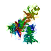
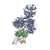
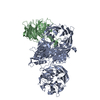
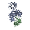
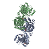
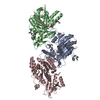
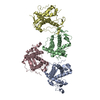
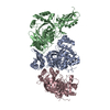
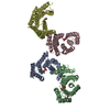
| Title | Structure of the hsDDB1-hsCSA complex | ||||||
 Components Components |
| ||||||
 Keywords Keywords |  DNA BINDING PROTEIN / DNA DAMAGE REPAIR DNA BINDING PROTEIN / DNA DAMAGE REPAIR | ||||||
| Function / homology |  Function and homology information Function and homology informationregulation of transcription-coupled nucleotide-excision repair / nucleotide-excision repair complex / single strand break repair / positive regulation by virus of viral protein levels in host cell / epigenetic programming in the zygotic pronuclei / spindle assembly involved in female meiosis / double-strand break repair via classical nonhomologous end joining / Cul4-RING E3 ubiquitin ligase complex / UV-damage excision repair / biological process involved in interaction with symbiont ...regulation of transcription-coupled nucleotide-excision repair / nucleotide-excision repair complex / single strand break repair / positive regulation by virus of viral protein levels in host cell / epigenetic programming in the zygotic pronuclei / spindle assembly involved in female meiosis / double-strand break repair via classical nonhomologous end joining / Cul4-RING E3 ubiquitin ligase complex / UV-damage excision repair / biological process involved in interaction with symbiont / regulation of mitotic cell cycle phase transition / WD40-repeat domain binding / Cul4A-RING E3 ubiquitin ligase complex / Cul4B-RING E3 ubiquitin ligase complex / ubiquitin ligase complex scaffold activity / negative regulation of reproductive process / negative regulation of developmental process / cullin family protein binding / response to X-ray / transcription-coupled nucleotide-excision repair / viral release from host cell / ectopic germ cell programmed cell death / protein autoubiquitination / positive regulation of viral genome replication / response to UV / positive regulation of gluconeogenesis / positive regulation of DNA repair / proteasomal protein catabolic process / nucleotide-excision repair / Recognition of DNA damage by PCNA-containing replication complex / DNA Damage Recognition in GG-NER / regulation of circadian rhythm / Dual Incision in GG-NER / Transcription-Coupled Nucleotide Excision Repair (TC-NER) / Formation of TC-NER Pre-Incision Complex / Wnt signaling pathway / nuclear matrix / Formation of Incision Complex in GG-NER / protein polyubiquitination / Dual incision in TC-NER / Gap-filling DNA repair synthesis and ligation in TC-NER / positive regulation of protein catabolic process / cellular response to UV / rhythmic process / protein-macromolecule adaptor activity / site of double-strand break / Neddylation / ubiquitin-dependent protein catabolic process / proteasome-mediated ubiquitin-dependent protein catabolic process / response to oxidative stress / chromosome, telomeric region / damaged DNA binding / protein ubiquitination / DNA repair / apoptotic process / DNA damage response / protein-containing complex binding / nucleolus / negative regulation of apoptotic process / protein-containing complex / DNA binding / extracellular space / extracellular exosome / nucleoplasm / nucleus / cytoplasmSimilarity search - Function | ||||||
| Biological species |  HOMO SAPIENS (human) HOMO SAPIENS (human) | ||||||
| Method | X-RAY DIFFRACTION / SYNCHROTRON / MOLECULAR REPLACEMENT / Resolution: 3.31 Å | ||||||
 Authors Authors | Bohm, K. / Scrima, A. / Fischer, E.S. / Gut, H. / Thomae, N.H. | ||||||
 Citation Citation | Journal: Cell(Cambridge,Mass.) / Year: 2011 Title: The Molecular Basis of Crl4(Ddb2/Csa) Ubiquitin Ligase Architecture, Targeting, and Activation. Authors: Fischer, E.S. / Scrima, A. / Bohm, K. / Matsumoto, S. / Lingaraju, G.M. / Faty, M. / Yasuda, T. / Cavadini, S. / Wakasugi, M. / Hanaoka, F. / Iwai, S. / Gut, H. / Sugasawa, K. / Thoma, N.H. | ||||||
| History |
|
- Structure visualization
| Structure viewer | Molecule: MolmilJmol/JSmol |
|---|
- Downloads & links
-Download
| PDBx/mmCIF format | 4a11.cif.gz | 568 KB | Display | PDBx/mmCIF format |
|---|---|---|---|---|
| PDB format | pdb4a11.ent.gz | 467.2 KB | Display | PDB format |
| PDBx/mmJSON format | 4a11.json.gz | Tree view | PDBx/mmJSON format | |
| Others |  Other downloads Other downloads |
-Validation report
| Arichive directory | https://data.pdbj.org/pub/pdb/validation_reports/a1/4a11ftp://data.pdbj.org/pub/pdb/validation_reports/a1/4a11 | HTTPS FTP |
|---|
-Related structure data
| Related structure data | 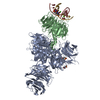 4a08C 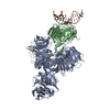 4a09C 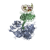 4a0aC 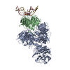 4a0bC 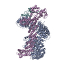 4a0cC 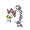 4a0kC 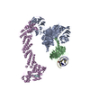 4a0lC 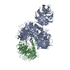 3ei3S C: citing same article ( S: Starting model for refinement |
|---|---|
| Similar structure data |


-Links
 PDBj
PDBj


- Assembly
| Deposited unit | 
| ||||||||
|---|---|---|---|---|---|---|---|---|---|
| 1 |
| ||||||||
| Unit cell |
|
-Components
| #1: Protein | Mass: 129394.898 Da / Num. of mol.: 1 Source method: isolated from a genetically manipulated source Source: (gene. exp.) HOMO SAPIENS (human) / Plasmid: PFASTBAC-DERIVED / Cell line (production host): High Five / Production host:  TRICHOPULSIA NI (cabbage looper) / References: UniProt: Q16531 TRICHOPULSIA NI (cabbage looper) / References: UniProt: Q16531 |
|---|---|
| #2: Protein | Mass: 45465.613 Da / Num. of mol.: 1 Source method: isolated from a genetically manipulated source Source: (gene. exp.) HOMO SAPIENS (human) / Plasmid: PFASTBAC-DERIVED / Cell line (production host): High Five / Production host: TRICHOPULSIA NI (cabbage looper) / References: UniProt: Q13216 |
-Experimental details
-Experiment
| Experiment | Method: X-RAY DIFFRACTION |
|---|
- Sample preparation
| Crystal | Density Matthews: 4.05 Å3/Da / Density % sol: 69 % / Description: NONE |
|---|---|
| Crystal grow | pH: 8 Details: 1.4-1.58 M NAKPO4, 0.1 M NAMALONATE, 0-0.1 M LI2SO4, pH 8.0 |
-Data collection
| Diffraction | Mean temperature: 100 K |
|---|---|
| Diffraction source | Source: SYNCHROTRON / Site: SLS  / Beamline: X10SA / Wavelength: 1 / Beamline: X10SA / Wavelength: 1 |
| Detector | Type: DECTRIS PILATUS 6M / Detector: PIXEL |
| Radiation | Protocol: SINGLE WAVELENGTH / Monochromatic (M) / Laue (L): M / Scattering type: x-ray |
| Radiation wavelength | Wavelength: 1 Å / Relative weight: 1 |
| Reflection | Resolution: 3.3→50 Å / Num. obs: 38124 / % possible obs: 92.4 % / Observed criterion σ(I): -3 / Redundancy: 2.2 % / Biso Wilson estimate: 80.81 Å2 / Rmerge(I) obs: 0.1 / Net I/σ(I): 7.03 |
| Reflection shell | Resolution: 3.31→3.39 Å / Redundancy: 1.9 % / Rmerge(I) obs: 0.43 / Mean I/σ(I) obs: 1.95 / % possible all: 78.4 |
- Processing
| Software |
| |||||||||||||||||||||||||||||||||||||||||||||||||||||||||||||||||||||||||||||||||||||||||||||||||||||||||
|---|---|---|---|---|---|---|---|---|---|---|---|---|---|---|---|---|---|---|---|---|---|---|---|---|---|---|---|---|---|---|---|---|---|---|---|---|---|---|---|---|---|---|---|---|---|---|---|---|---|---|---|---|---|---|---|---|---|---|---|---|---|---|---|---|---|---|---|---|---|---|---|---|---|---|---|---|---|---|---|---|---|---|---|---|---|---|---|---|---|---|---|---|---|---|---|---|---|---|---|---|---|---|---|---|---|---|
| Refinement | Method to determine structure: MOLECULAR REPLACEMENT Starting model: PDB ENTRY 3EI3 Resolution: 3.31→32.067 Å / SU ML: 0.69 / σ(F): 1.99 / Phase error: 21.77 / Stereochemistry target values: ML Details: UNMODELLED DENSITY WAS OBSERVED CLOSE THE THE DDB1-BPB DOMAIN POTENTIALLY REFLECTING A MAINLY UNSTRUCTURED LOOP REGION OF DDB1.
| |||||||||||||||||||||||||||||||||||||||||||||||||||||||||||||||||||||||||||||||||||||||||||||||||||||||||
| Solvent computation | Shrinkage radii: 0.83 Å / VDW probe radii: 1.1 Å / Solvent model: FLAT BULK SOLVENT MODEL / Bsol: 74.497 Å2 / ksol: 0.326 e/Å3 | |||||||||||||||||||||||||||||||||||||||||||||||||||||||||||||||||||||||||||||||||||||||||||||||||||||||||
| Displacement parameters | Biso mean: 84.02 Å2
| |||||||||||||||||||||||||||||||||||||||||||||||||||||||||||||||||||||||||||||||||||||||||||||||||||||||||
| Refinement step | Cycle: LAST / Resolution: 3.31→32.067 Å
| |||||||||||||||||||||||||||||||||||||||||||||||||||||||||||||||||||||||||||||||||||||||||||||||||||||||||
| Refine LS restraints |
| |||||||||||||||||||||||||||||||||||||||||||||||||||||||||||||||||||||||||||||||||||||||||||||||||||||||||
| LS refinement shell |
|
