 Movie
Movie Controller

[English] 日本語
 Yorodumi
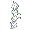
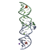
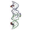
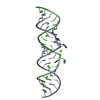
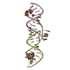
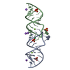
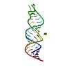
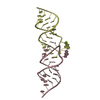
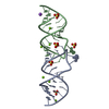
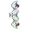
- PDB-462d: CRYSTAL STRUCTURE OF THE HIV-1 GENOMIC RNA DIMERIZATION INITIATIO... -
+ Open data

- Basic information
| Entry | Database: PDB / ID: 462d | ||||||||||||||||||
|---|---|---|---|---|---|---|---|---|---|---|---|---|---|---|---|---|---|---|---|
| Title | CRYSTAL STRUCTURE OF THE HIV-1 GENOMIC RNA DIMERIZATION INITIATION SITE | ||||||||||||||||||
 Components Components | RNA (5'-R(* Keywords Keywords RNA / HIV / RNA DUPLEX / MISMATCH / BULGES / MAGNESIUM BINDING RNA / HIV / RNA DUPLEX / MISMATCH / BULGES / MAGNESIUM BINDINGFunction / homology | RNA / RNA (> 10) Function and homology information Function and homology informationMethod | X-RAY DIFFRACTION / SYNCHROTRON / MIRAS / Resolution: 2.3 Å  Authors AuthorsEnnifar, E. / Yusupov, M. / Walter, P. / Marquet, R. / Ehresmann, C. / Ehresmann, B. / Dumas, P. |  CitationJournal: Structure Fold.Des. / Year: 1999 CitationJournal: Structure Fold.Des. / Year: 1999Title: The crystal structure of the dimerization initiation site of genomic HIV-1 RNA reveals an extended duplex with two adenine bulges. Authors: Ennifar, E. / Yusupov, M. / Walter, P. / Marquet, R. / Ehresmann, B. / Ehresmann, C. / Dumas, P. History |
|
- Structure visualization
| Structure viewer | Molecule: MolmilJmol/JSmol |
|---|
- Downloads & links
-Download
| PDBx/mmCIF format | 462d.cif.gz | 45.4 KB | Display | PDBx/mmCIF format |
|---|---|---|---|---|
| PDB format | pdb462d.ent.gz | 32 KB | Display | PDB format |
| PDBx/mmJSON format | 462d.json.gz | Tree view | PDBx/mmJSON format | |
| Others |  Other downloads Other downloads |
-Validation report
| Arichive directory | https://data.pdbj.org/pub/pdb/validation_reports/62/462dftp://data.pdbj.org/pub/pdb/validation_reports/62/462d | HTTPS FTP |
|---|
-Related structure data


-Links
 PDBj
PDBj


- Assembly
| Deposited unit | 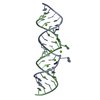
| |||||||||||||||
|---|---|---|---|---|---|---|---|---|---|---|---|---|---|---|---|---|
| 1 |
| |||||||||||||||
| Unit cell |
| |||||||||||||||
| Components on special symmetry positions |
| |||||||||||||||
| Noncrystallographic symmetry (NCS) | NCS oper: (Code: given Matrix: (-0.9998, 0.0196, 0.0093), Vector : |
-Components
| #1: RNA chain | Mass: 7402.472 Da / Num. of mol.: 2 / Source method: obtained synthetically #2: Chemical | ChemComp-MG /   Mass: 24.305 Da / Num. of mol.: 8 / Source method: obtained synthetically / Formula: Mg Mass: 24.305 Da / Num. of mol.: 8 / Source method: obtained synthetically / Formula: Mg#3: Water | ChemComp-HOH / | Water Mass: 18.015 Da / Num. of mol.: 240 / Source method: isolated from a natural source / Formula: H2O Mass: 18.015 Da / Num. of mol.: 240 / Source method: isolated from a natural source / Formula: H2O |
|---|
-Experimental details
-Experiment
| Experiment | Method: X-RAY DIFFRACTION / Number of used crystals: 2 |
|---|
- Sample preparation
| Crystal | Density Matthews: 2.18 Å3/Da / Density % sol: 50 % | ||||||||||||||||||||||||||||||||||||||||||||||||||||||||||||||||||
|---|---|---|---|---|---|---|---|---|---|---|---|---|---|---|---|---|---|---|---|---|---|---|---|---|---|---|---|---|---|---|---|---|---|---|---|---|---|---|---|---|---|---|---|---|---|---|---|---|---|---|---|---|---|---|---|---|---|---|---|---|---|---|---|---|---|---|---|
| Crystal grow | Temperature: 310 K / Method: vapor diffusion, sitting drop / pH: 7 Details: DROP: CACODYLATE 25MM PH 7.0, MGCL2 5MM, KCL 150MM, MPD 1%, SPERMINE 5MM; RESERVOIR: CACODYLATE 50MM PH 7.0, MGCL2 100MM, KCL 300MM, MPD 50%, VAPOR DIFFUSION, SITTING DROP, temperature 310K | ||||||||||||||||||||||||||||||||||||||||||||||||||||||||||||||||||
| Components of the solutions |
| ||||||||||||||||||||||||||||||||||||||||||||||||||||||||||||||||||
| Crystal grow | *PLUS Temperature: 37 ℃ / Method: vapor diffusion | ||||||||||||||||||||||||||||||||||||||||||||||||||||||||||||||||||
| Components of the solutions | *PLUS
|
-Data collection
| Diffraction |
| |||||||||||||||||||||
|---|---|---|---|---|---|---|---|---|---|---|---|---|---|---|---|---|---|---|---|---|---|---|
| Diffraction source |
| |||||||||||||||||||||
| Detector |
| |||||||||||||||||||||
| Radiation |
| |||||||||||||||||||||
| Radiation wavelength |
| |||||||||||||||||||||
| Reflection | Resolution: 2.3→15 Å / Num. all: 5842 / Num. obs: 5842 / % possible obs: 97.9 % / Observed criterion σ(I): 0 / Redundancy: 6.5 % / Biso Wilson estimate: 43 Å2 / Rmerge(I) obs: 0.069 | |||||||||||||||||||||
| Reflection shell | Resolution: 2.3→2.38 Å / Redundancy: 6 % / Rmerge(I) obs: 0.135 / % possible all: 98.3 | |||||||||||||||||||||
| Reflection | *PLUS Highest resolution: 2.25 Å | |||||||||||||||||||||
| Reflection shell | *PLUS % possible obs: 98.3 % |

- Processing
| Software |
| ||||||||||||||||||||||||||||||||||||||||||||||||||||||||||||
|---|---|---|---|---|---|---|---|---|---|---|---|---|---|---|---|---|---|---|---|---|---|---|---|---|---|---|---|---|---|---|---|---|---|---|---|---|---|---|---|---|---|---|---|---|---|---|---|---|---|---|---|---|---|---|---|---|---|---|---|---|---|
| Refinement | Method to determine structure: MIRAS / Resolution: 2.3→10 Å / Rfactor Rfree error: 0.008 / Data cutoff high absF: 1942623.23 / Data cutoff low absF: 0 / Isotropic thermal model: RESTRAINED / Cross valid method: THROUGHOUT / σ(F): 0 Details: DERIVATIVE TO NATIVE SCALING AND HEAVY ATOM SEARCH DONE WITH PROGRAM LOCHVAT ( DUMAS, 1994, ACTA. CRYST. A50, 526-534, 537-546)
| ||||||||||||||||||||||||||||||||||||||||||||||||||||||||||||
| Solvent computation | Solvent model: FLAT MODEL / Bsol: 63.97 Å2 / ksol: 0.33 e/Å3 | ||||||||||||||||||||||||||||||||||||||||||||||||||||||||||||
| Displacement parameters | Biso mean: 44.1 Å2
| ||||||||||||||||||||||||||||||||||||||||||||||||||||||||||||
| Refine analyze |
| ||||||||||||||||||||||||||||||||||||||||||||||||||||||||||||
| Refinement step | Cycle: LAST / Resolution: 2.3→10 Å
| ||||||||||||||||||||||||||||||||||||||||||||||||||||||||||||
| Refine LS restraints |
| ||||||||||||||||||||||||||||||||||||||||||||||||||||||||||||
| LS refinement shell | Resolution: 2.3→2.44 Å / Rfactor Rfree error: 0.036 / Total num. of bins used: 6
| ||||||||||||||||||||||||||||||||||||||||||||||||||||||||||||
| Xplor file |
| ||||||||||||||||||||||||||||||||||||||||||||||||||||||||||||
| Software | *PLUS Name: CNS / Version: 0.4 / Classification: refinement | ||||||||||||||||||||||||||||||||||||||||||||||||||||||||||||
| Refine LS restraints | *PLUS
|
